00 Foundations
Kramers bremsstrahlung spectrum
Unfiltered thick-target intensity I(E) ∝ Z (E_max − E), E_max = kVp.
Listen
Listen · English
Simulation
Kramers bremsstrahlung spectrum — Change the numbers; the scene follows.
Where it works
Radiography room
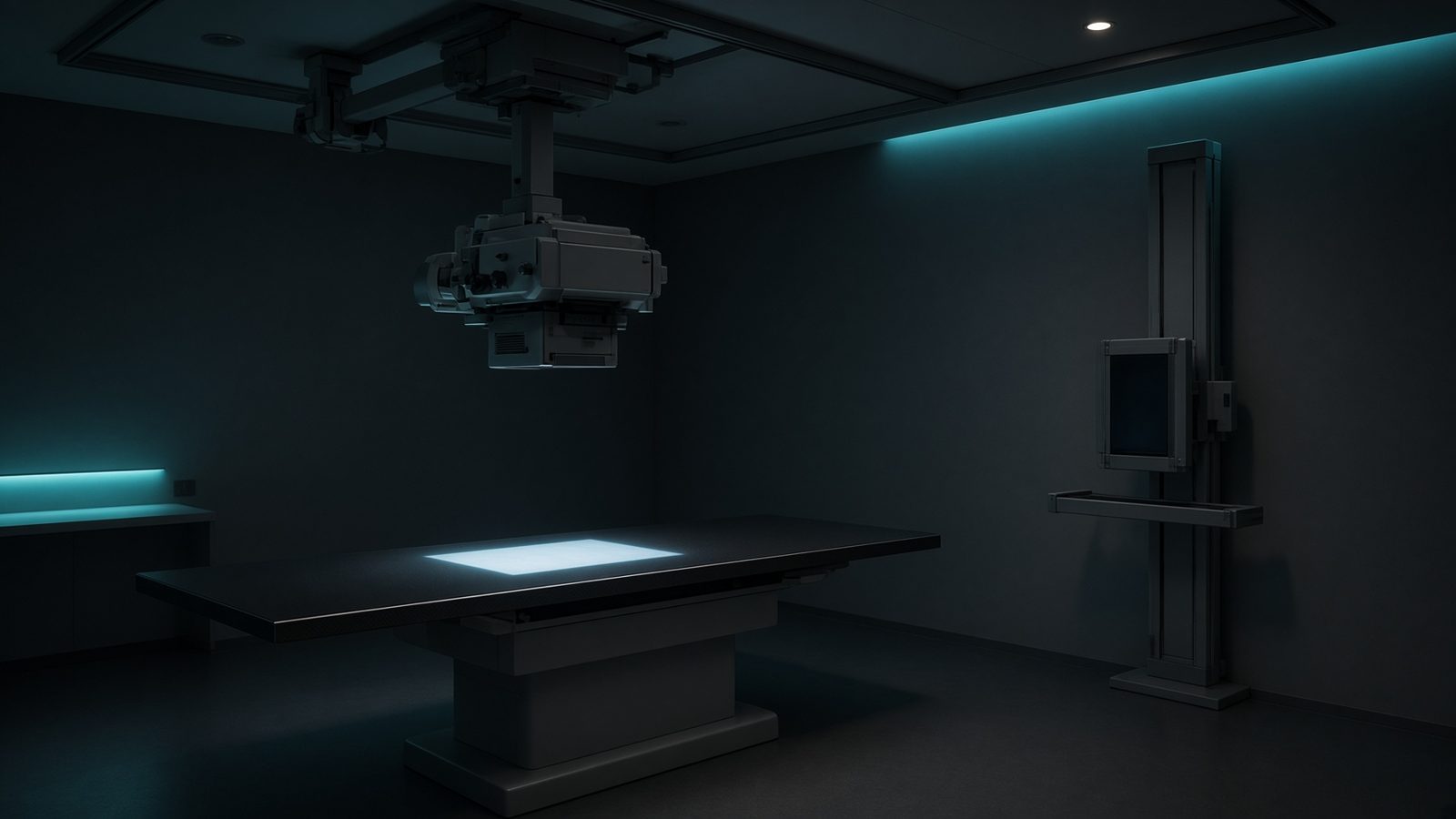
X-ray tube
At the focal spot in the tube housing — spectrum, output, SID geometry, and unsharpness start here.
Open this machineFormula
Variables
Results
I(E)
Relative intensity
2,960
I/I(0)
Fraction of I(0)
0.5
Ē
Unfiltered mean energy
26.6667keV
E_eff
Rough filtered effective energy
40keV
Curve
Explanation
What it means
In a thick anode the electron slows from e·kVp to rest, radiating a triangular photon spectrum that is maximum at 0 keV and zero at E_max. Filtration (inherent + added Al) cuts the low-energy end, so a clinical beam peaks near E_max/2 to E_max/3 and the mean energy is ~E_max/3 to ~E_max/2. Tube output scales as Z of the anode and roughly as kVp² (after filtration, closer to kVp²–kVp³). This is a working relation in Radiation physics.
Where it is used
Clinically it sits on the Radiography room — X-ray tube. At the focal spot in the tube housing — spectrum, output, SID geometry, and unsharpness start here. Radiation physics lives at the x-ray target, the linac head, and inside the patient: how a photon is born, how it scatters, and how it dies. Use these relations before you trust a spectrum, a wall, or a kV-versus-MV contrast argument.
Radiography room · Open this machineHow to use it
Enter kVp, anode Z, and the energy E at which you want relative intensity. The curve shows the unfiltered Kramers triangle. Use it to explain why 120 kVp CT has useful flux up to 120 keV and why 25 kVp mammography needs a Mo/Rh spectrum instead. Change one input and watch the curve and the simulation follow.
Symbols
- kVpTube voltage80 kV
- ZAnode Z74
- EPhoton energy40 keV
Worked example
A typical case from the default values: kVp = 80 kV (Tube voltage); Z = 74 (Anode Z); E = 40 keV (Photon energy). Substituting into the relation gives I(E) = 2,960; I/I(0) = 0.5; Ē = 26.6667 keV; E_eff = 40 keV. These are teaching numbers — align them with your machine.
Typical values give
- I(E) = 2,960
- I/I(0) = 0.5
- Ē = 26.6667keV
- E_eff = 40keV
Where it comes from
The displayed formula is the working relation. Unfiltered thick-target intensity I(E) ∝ Z (E_max − E), E_max = kVp. Usual reference: Kramers 1923 / Bushberg. Derive it in the specialty lesson, then return here to pin the numbers.
Reference: Kramers 1923 / Bushberg
Assumptions & limits
No filtration, no characteristic lines, no heel effect, no off-axis hardening. Real spectra are the Kramers triangle × exp(−μ(E)x) plus K and L lines sitting on top.
Pitfalls
Do not mix free-electron Compton kinematics with photoelectric-dominated kV imaging. Check keV versus MeV, and never treat a spectrum as one photon. No filtration, no characteristic lines, no heel effect, no off-axis hardening. Real spectra are the Kramers triangle × exp(−μ(E)x) plus K and L lines sitting on top.
Keep this
Photons do not deposit dose; the electrons they set in motion do. No filtration, no characteristic lines, no heel effect, no off-axis hardening. Real spectra are the Kramers triangle × exp(−μ(E)x) plus K and L lines sitting on top.
In this specialty