Lecture
Library
A lecture cinema of its own — specialty films and machine films, with voice, a laser you can hide, and a notebook.
9 Specialty films · 13 Machine films
Specialty films
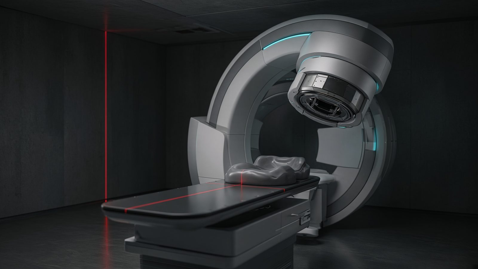 8 min00 Foundations
How a photon dies in tissue
Three ways a photon can vanish — and why the one that wins depends on energy and atomic number.
EN FR AR ES
- 9 min01 Therapy
From isocenter to monitor units
Inverse square, percent depth dose, and the MU you will actually type.
EN FR AR ES
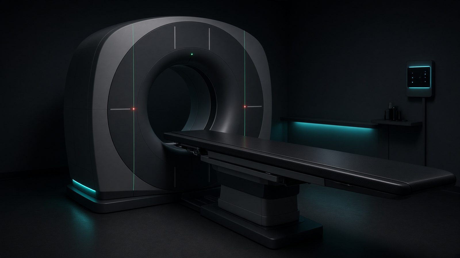 8 min02 Imaging
Dose indices you will be asked
CTDI, DLP, SSDE, and why a single CTDI is not the patient's dose.
EN FR AR ES
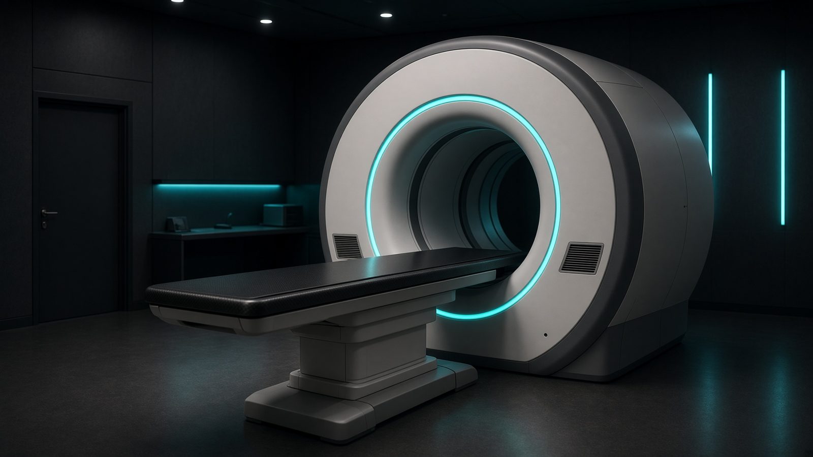 8 min03 Nuclear
Activity, wait, and the number on the report
Decay is not a nuisance. It is the clock of every elution, every SUV, every delayed image.
EN FR AR ES
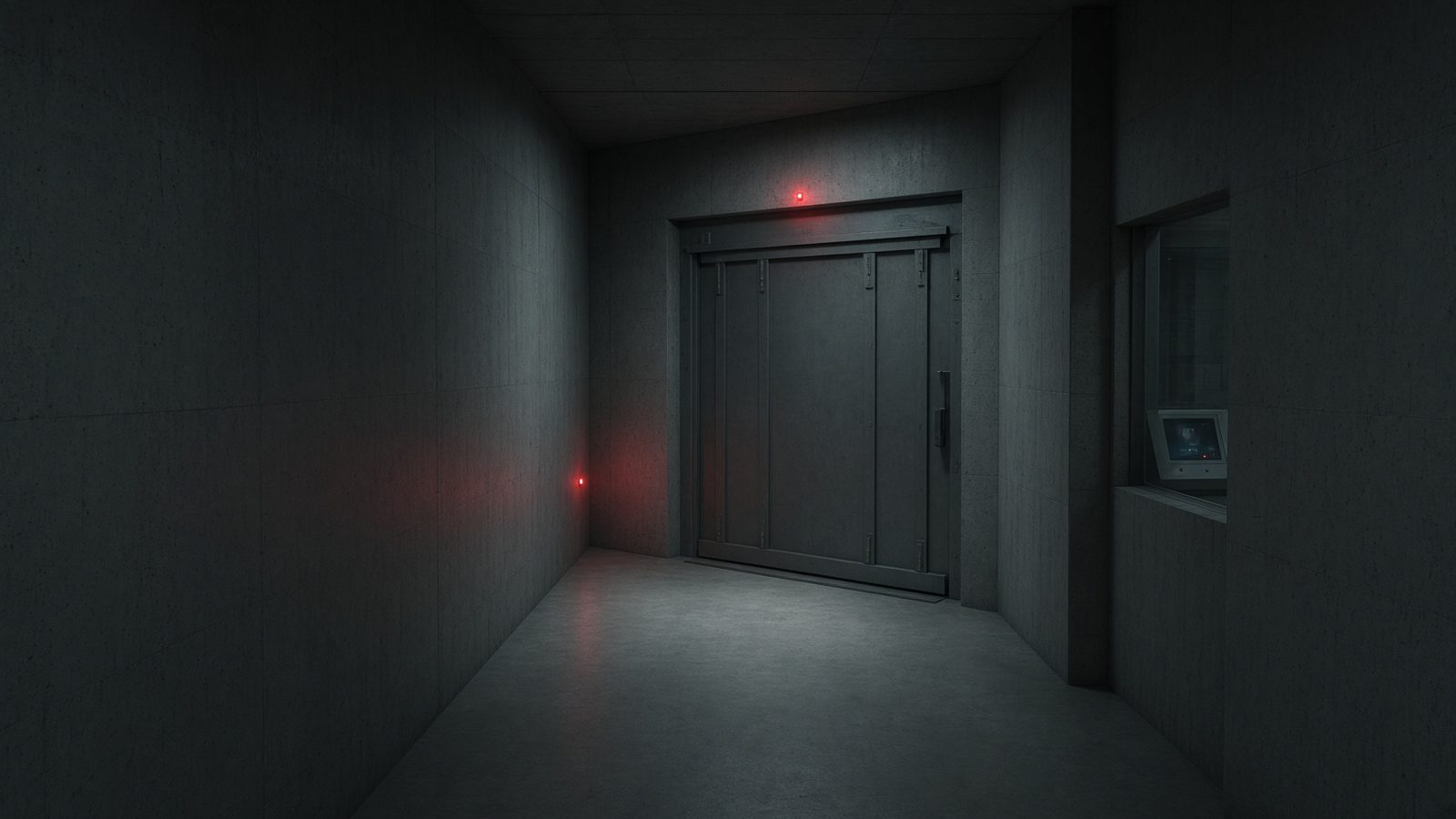 7 min04 Protection
Time, distance, shielding — then the wall
The three controls you already know, written so a barrier calculation has a place to start.
EN FR AR ES
- 8 min05 Biology
Fractionation is a language
Linear-quadratic survival, BED, and EQD2 — how a 2-gray fraction and a 8-gray fraction stop being comparable until you convert.
EN FR AR ES
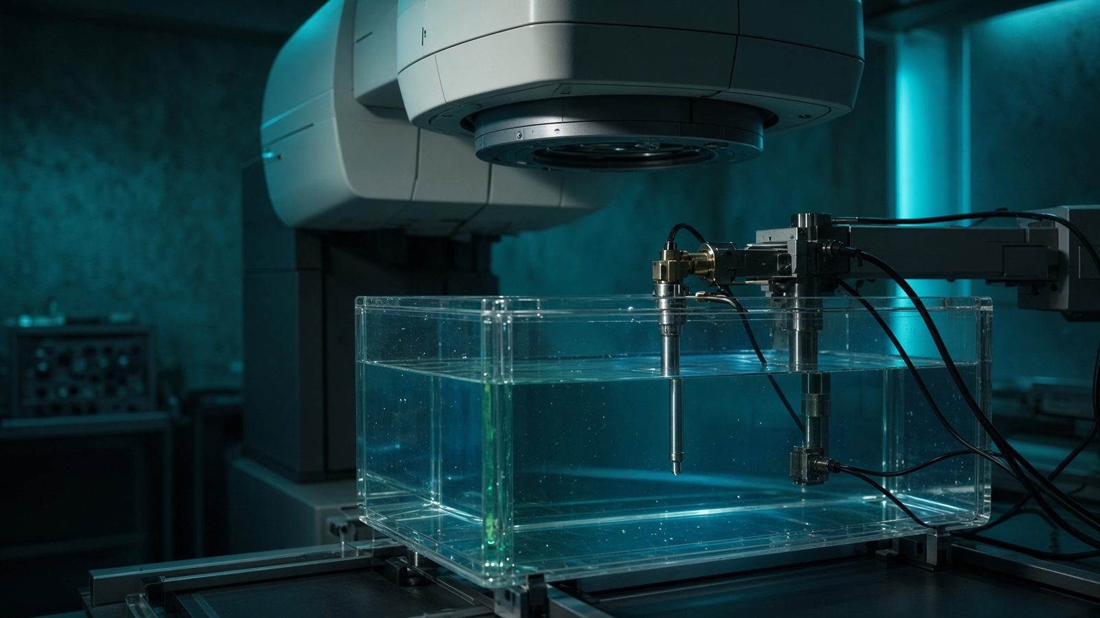 8 min06 Dose
From charge in a cavity to gray in water
Bragg–Gray, TG-51, kQ and kTP — the chain that turns a coulomb into absorbed dose.
EN FR AR ES
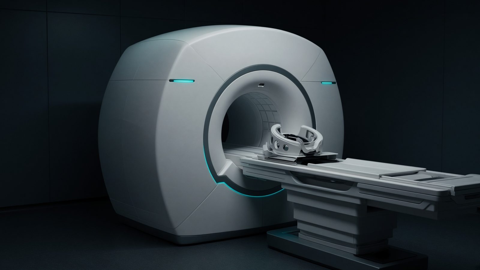 8 min07 MRI
A clock, a tip, a recovery
Larmor frequency, the RF tip, and T1/T2 — contrast is a timing choice.
EN FR AR ES
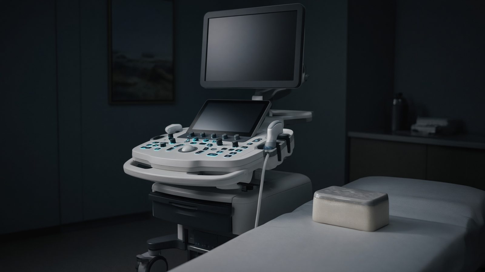 7 min08 Ultrasound
A click, an echo, a shift
Impedance sends the echo home. Time is depth. Motion is Doppler. That is the whole craft.
EN FR AR ES
Machine films
- 4 min01 Therapy
Linear accelerator
C-arm linac in the bunker — monitor chamber, MLC, and the beam at isocenter.
EN FR AR ES
- 4 min02 Imaging
CT scanner
Diagnostic CT — tube and detectors in the gantry, dose along the scanned length.
EN FR AR ES
- 4 min07 MRI
MRI scanner
Wide-bore magnet — B₀, gradients, and the RF coil on the patient.
EN FR AR ES
- 4 min03 Nuclear
PET/CT
Hybrid scanner — coincidence ring behind, CT for attenuation in front.
EN FR AR ES
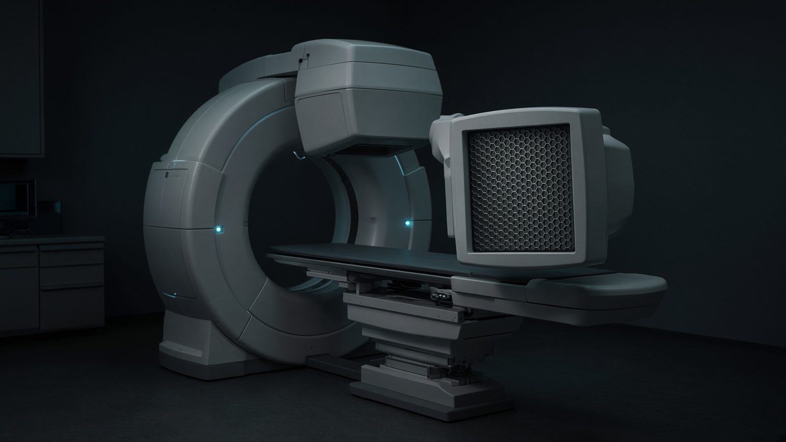 4 min03 Nuclear
Gamma camera
Dual-head SPECT — crystal, PMTs, and the collimator facing the patient.
EN FR AR ES
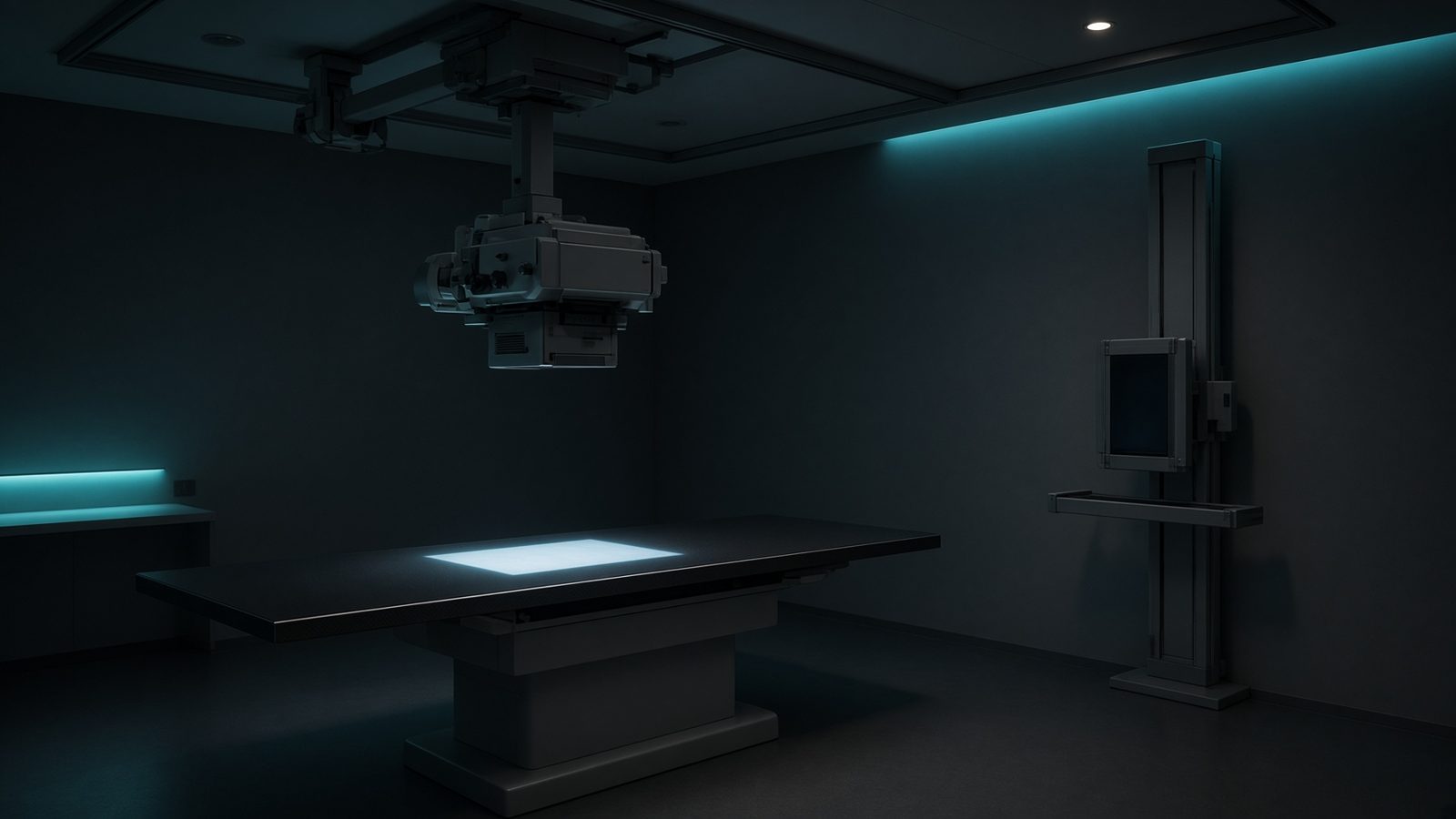 4 min02 Imaging
Radiography room
Ceiling tube, table, and wall Bucky — geometry, technique, and detector dose.
EN FR AR ES
- 4 min08 Ultrasound
Ultrasound
Probe on tissue — impedance, Doppler, and the console that sets PRF and MI.
EN FR AR ES
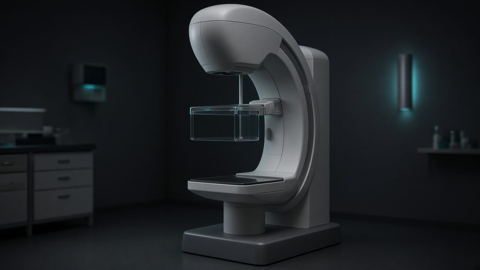 4 min02 Imaging
Mammography unit
C-arm with compression paddle — target/filter and average glandular dose.
EN FR AR ES
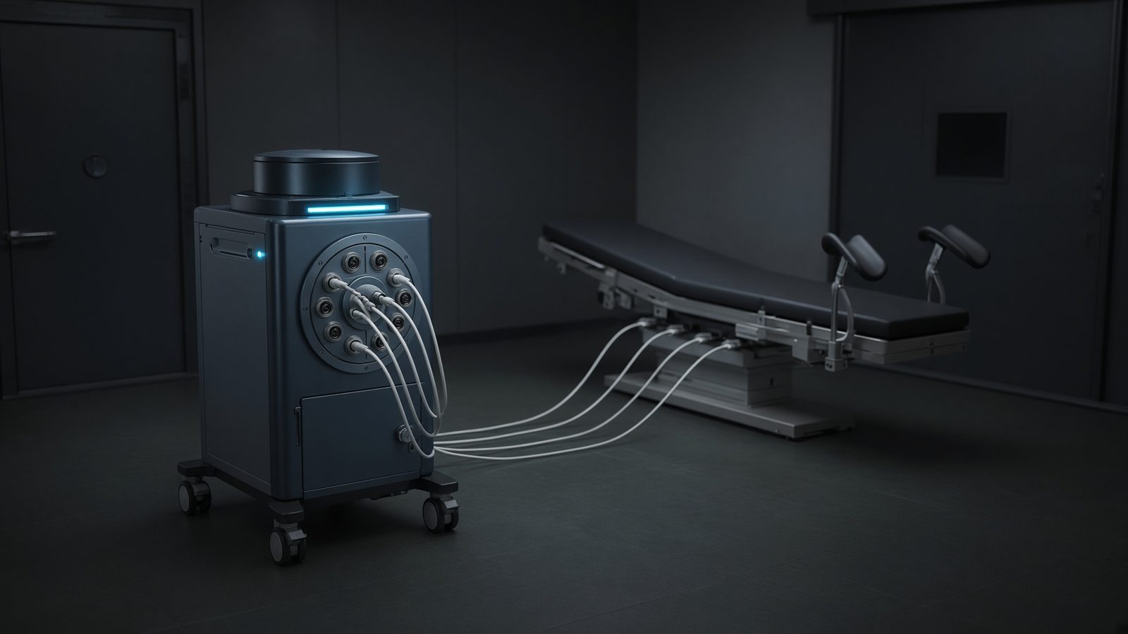 4 min01 Therapy
Brachytherapy suite
HDR afterloader and C-arm — source in the safe, dwell in the applicator.
EN FR AR ES
- 4 min06 Dose
Water phantom
Scanning tank under the linac — TG-51 / TRS-398 reference dosimetry.
EN FR AR ES
- 4 min04 Protection
Treatment vault
Maze and barriers — time, distance, shielding, WUT, and weekly dose.
EN FR AR ES
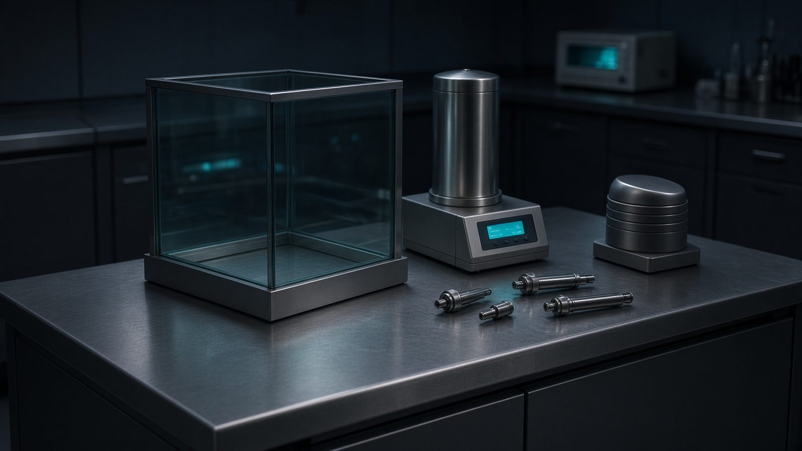 4 min03 Nuclear
Hot lab
Generator, dose calibrator, and L-block — activity, breakthrough, Marinelli.
EN FR AR ES
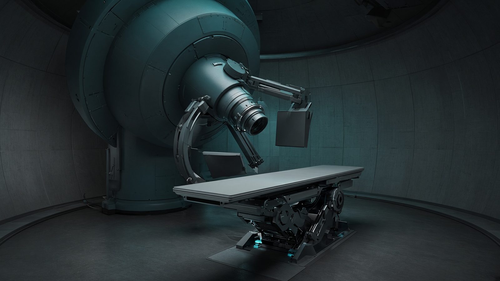 4 min01 Therapy
Proton gantry
Nozzle and snout — residual range, RBE 1.1, and the Bragg peak in the patient.
EN FR AR ES