Clinical lab
Physica
Medical physics equation lab
Dr. Tarik ELASRI
Founder · ASRIMP
Association for Scientific Research & Innovation in Medical Physics
www.asrimp.comA full equations library by specialty — then machines, lessons, and lecture films. Enter the numbers, watch the schematic, read a taught explanation.
Open the equations library216 equations · 9 specialties
Equations
See allA catalog of its own — every working relation by specialty, with the formula, a live calculator, and a taught explanation.
Radiation physics
Photons, Compton, Klein–Nishina, pair production, proton range, and activity.
22 eq.
Radiotherapy
Monitor units, PDD, TMR, electrons, TG-43, margins, and plan indices.
33 eq.
Diagnostic imaging
Magnification, CTDI, SSDE, DQE, AGD, grids, and technique rules.
29 eq.
Nuclear medicine
Decay, SUV, TOF, NEC, generators, counting stats, and cameras.
28 eq.
Radiation protection
Time, distance, shielding, WUT, equivalent dose, and barriers.
20 eq.
Radiobiology
LQ model, BED, EQD2, LKB, EUD, and TCP/NTCP.
20 eq.
Dosimetry
TG-51, TRS-398, kerma, Bragg-Gray, f-factor, and recombination.
20 eq.
MRI physics
Larmor, Ernst, diffusion, VENC, SAR, chemical shift, and relaxivity.
23 eq.
Ultrasound
Impedance, Doppler, MI, TI, Snell, PRF, and resolution.
21 eq.
Lessons
See all- 00 FoundationsHow a photon dies in tissueThree ways a photon can vanish — and why the one that wins depends on energy and atomic number.8 min · EN FR AR ES
- 01 TherapyFrom isocenter to monitor unitsInverse square, percent depth dose, and the MU you will actually type.9 min · EN FR AR ES
- 02 ImagingDose indices you will be askedCTDI, DLP, SSDE, and why a single CTDI is not the patient's dose.8 min · EN FR AR ES
- 03 NuclearActivity, wait, and the number on the reportDecay is not a nuisance. It is the clock of every elution, every SUV, every delayed image.8 min · EN FR AR ES
- 04 ProtectionTime, distance, shielding — then the wallThe three controls you already know, written so a barrier calculation has a place to start.7 min · EN FR AR ES
- 05 BiologyFractionation is a languageLinear-quadratic survival, BED, and EQD2 — how a 2-gray fraction and a 8-gray fraction stop being comparable until you convert.8 min · EN FR AR ES
- 06 DoseFrom charge in a cavity to gray in waterBragg–Gray, TG-51, kQ and kTP — the chain that turns a coulomb into absorbed dose.8 min · EN FR AR ES
- 07 MRIA clock, a tip, a recoveryLarmor frequency, the RF tip, and T1/T2 — contrast is a timing choice.8 min · EN FR AR ES
- 08 UltrasoundA click, an echo, a shiftImpedance sends the echo home. Time is depth. Motion is Doppler. That is the whole craft.7 min · EN FR AR ES
Library
See allA lecture cinema of its own — specialty films and machine films, with voice, a laser you can hide, and a notebook.
- 00 FoundationsHow a photon dies in tissueThree ways a photon can vanish — and why the one that wins depends on energy and atomic number.8 min · Lecture
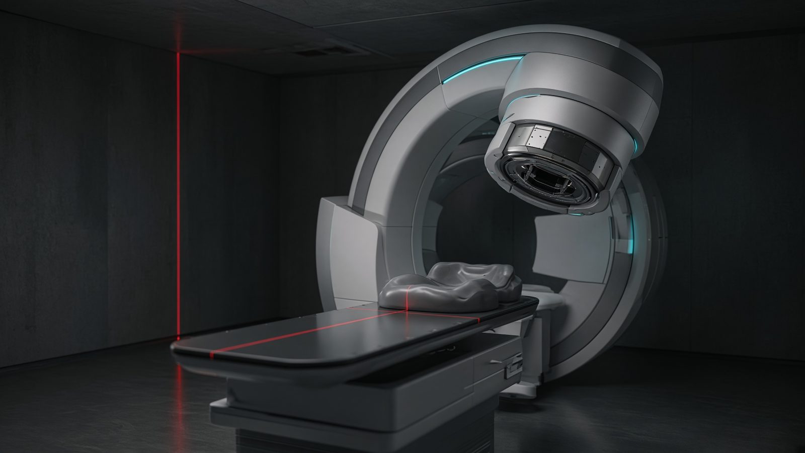
- 01 TherapyFrom isocenter to monitor unitsInverse square, percent depth dose, and the MU you will actually type.9 min · Lecture
- 02 ImagingDose indices you will be askedCTDI, DLP, SSDE, and why a single CTDI is not the patient's dose.8 min · Lecture
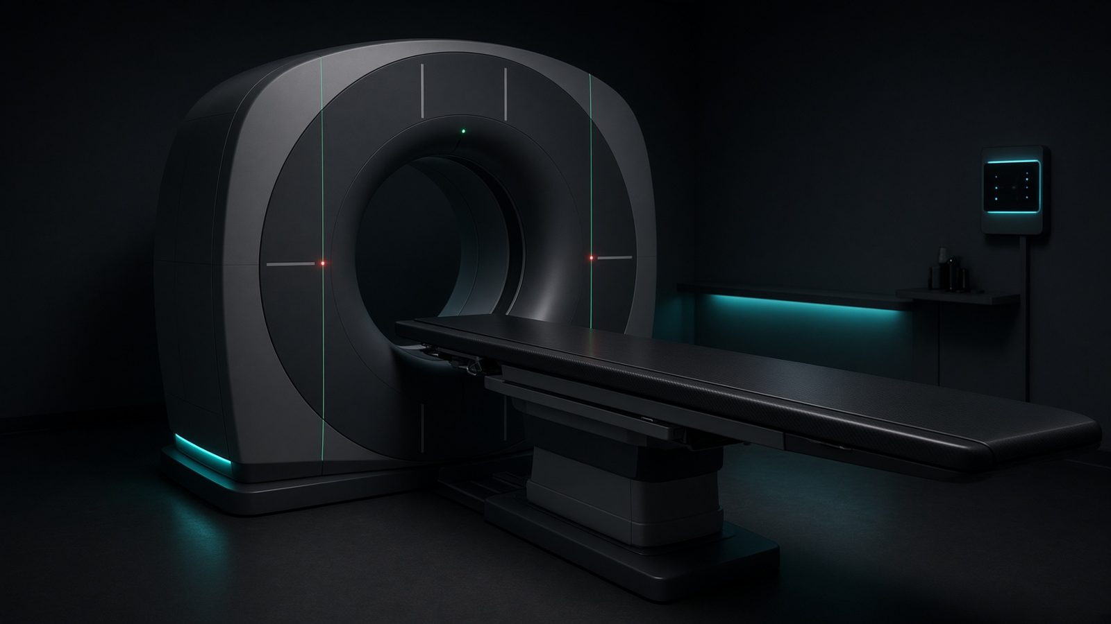
- 03 NuclearActivity, wait, and the number on the reportDecay is not a nuisance. It is the clock of every elution, every SUV, every delayed image.8 min · Lecture
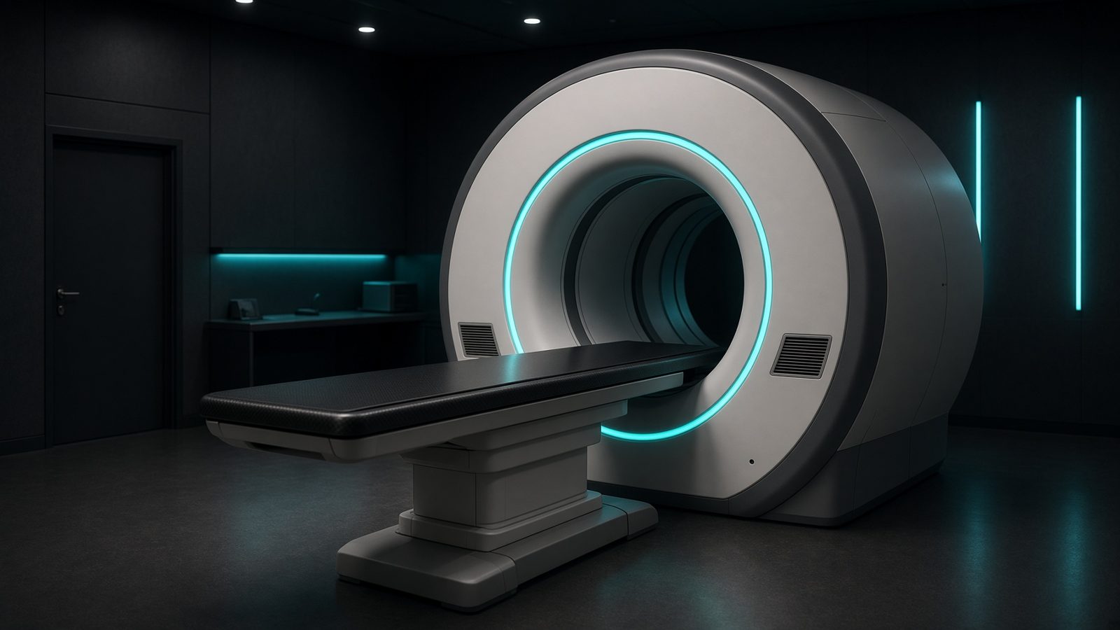
- 04 ProtectionTime, distance, shielding — then the wallThe three controls you already know, written so a barrier calculation has a place to start.7 min · Lecture
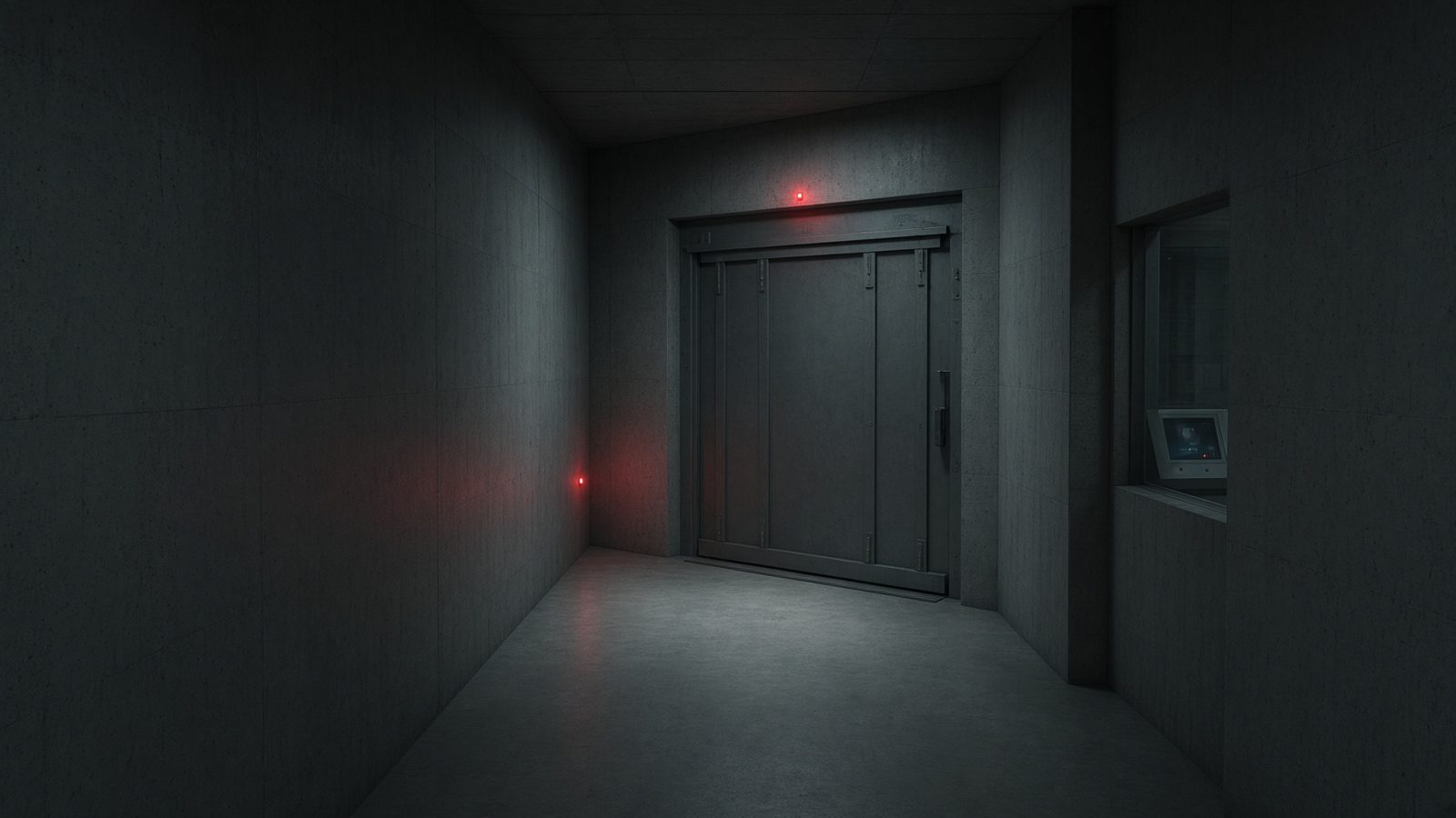
- 05 BiologyFractionation is a languageLinear-quadratic survival, BED, and EQD2 — how a 2-gray fraction and a 8-gray fraction stop being comparable until you convert.8 min · Lecture
- 06 DoseFrom charge in a cavity to gray in waterBragg–Gray, TG-51, kQ and kTP — the chain that turns a coulomb into absorbed dose.8 min · Lecture
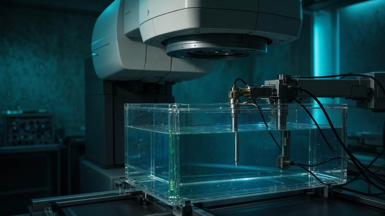
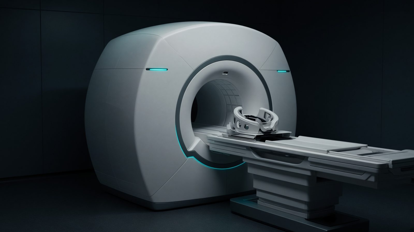
- 07 MRIA clock, a tip, a recoveryLarmor frequency, the RF tip, and T1/T2 — contrast is a timing choice.8 min · Lecture
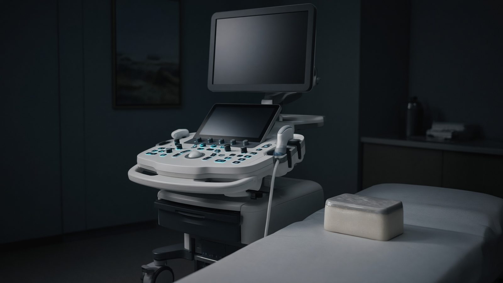
- 08 UltrasoundA click, an echo, a shiftImpedance sends the echo home. Time is depth. Motion is Doppler. That is the whole craft.7 min · Lecture
Machines
See all- Linear acceleratorC-arm linac in the bunker — monitor chamber, MLC, and the beam at isocenter.58 eq.
- CT scannerDiagnostic CT — tube and detectors in the gantry, dose along the scanned length.7 eq.
- MRI scannerWide-bore magnet — B₀, gradients, and the RF coil on the patient.23 eq.
- PET/CTHybrid scanner — coincidence ring behind, CT for attenuation in front.16 eq.
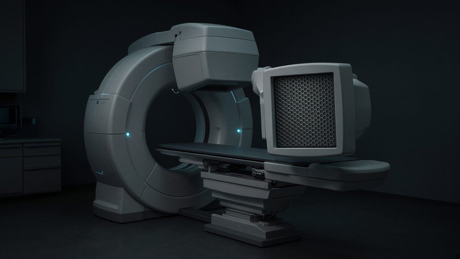 Gamma cameraDual-head SPECT — crystal, PMTs, and the collimator facing the patient.6 eq.
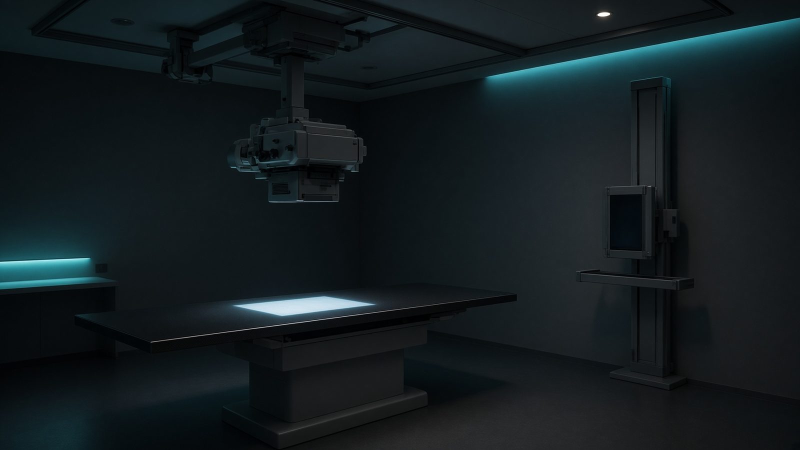 Radiography roomCeiling tube, table, and wall Bucky — geometry, technique, and detector dose.25 eq.
- UltrasoundProbe on tissue — impedance, Doppler, and the console that sets PRF and MI.21 eq.
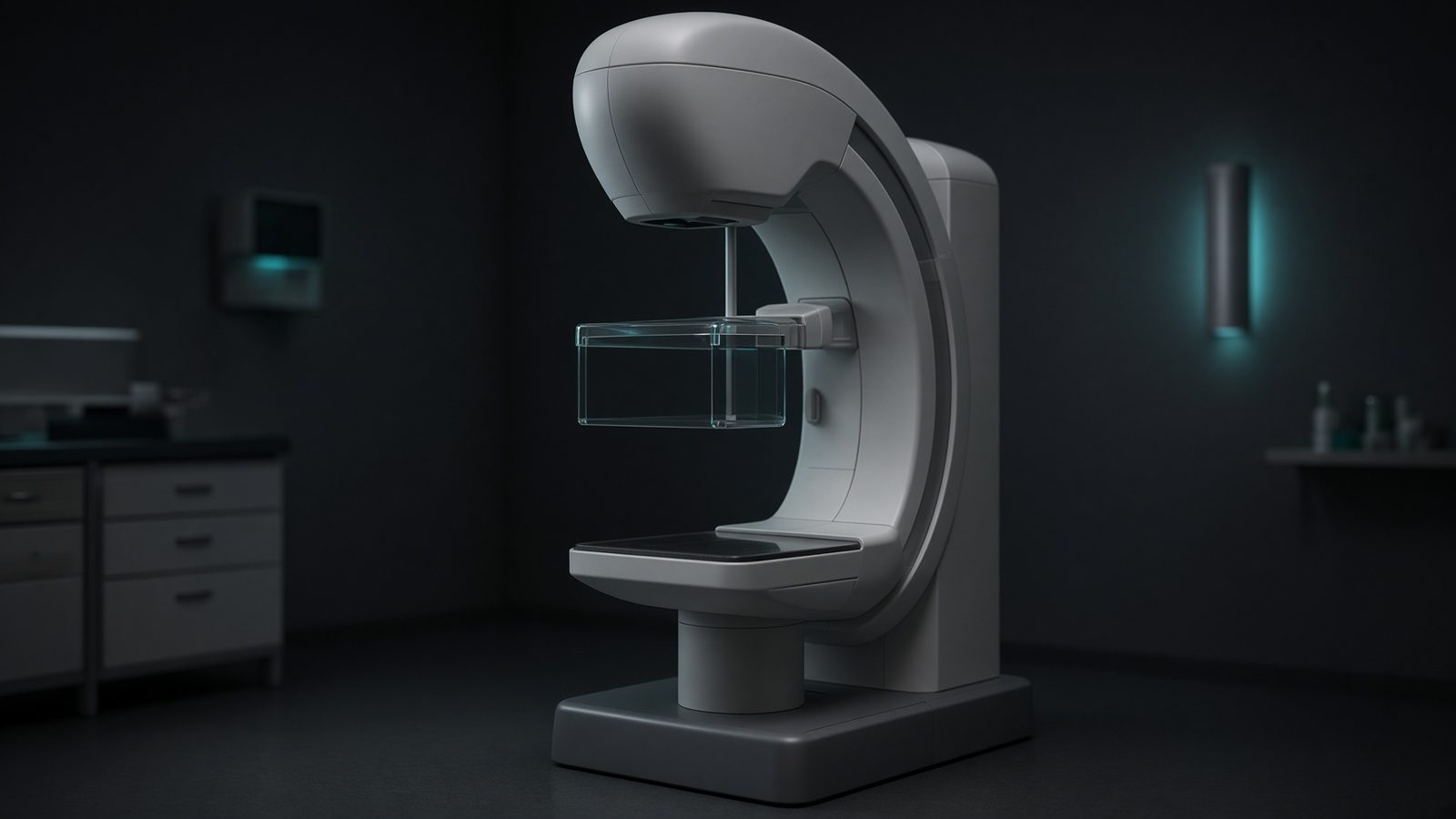 Mammography unitC-arm with compression paddle — target/filter and average glandular dose.1 eq.
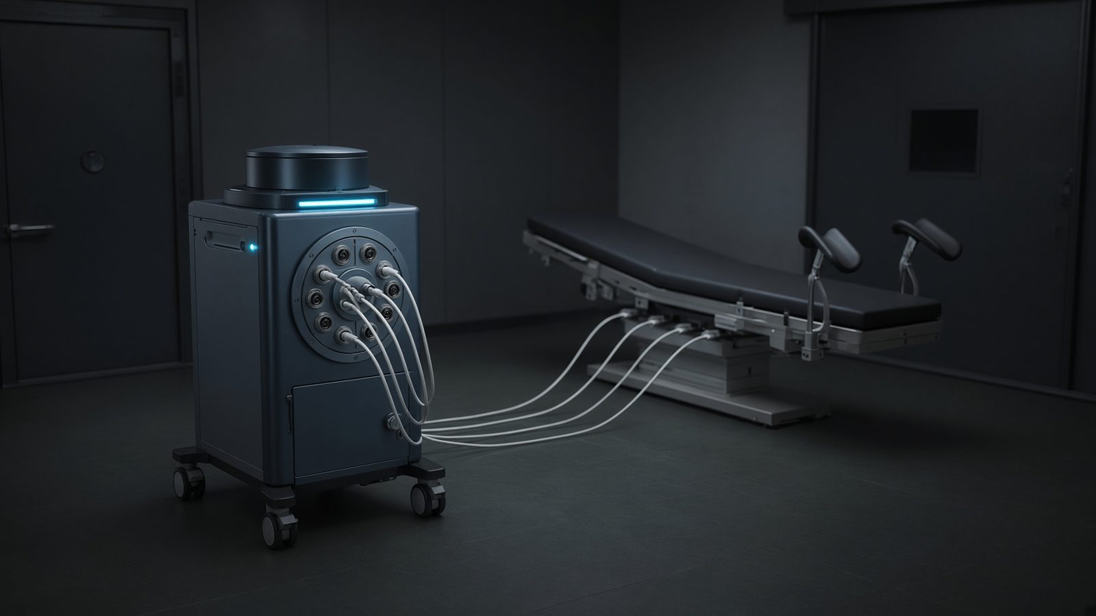 Brachytherapy suiteHDR afterloader and C-arm — source in the safe, dwell in the applicator.4 eq.
- Water phantomScanning tank under the linac — TG-51 / TRS-398 reference dosimetry.20 eq.
- Treatment vaultMaze and barriers — time, distance, shielding, WUT, and weekly dose.18 eq.
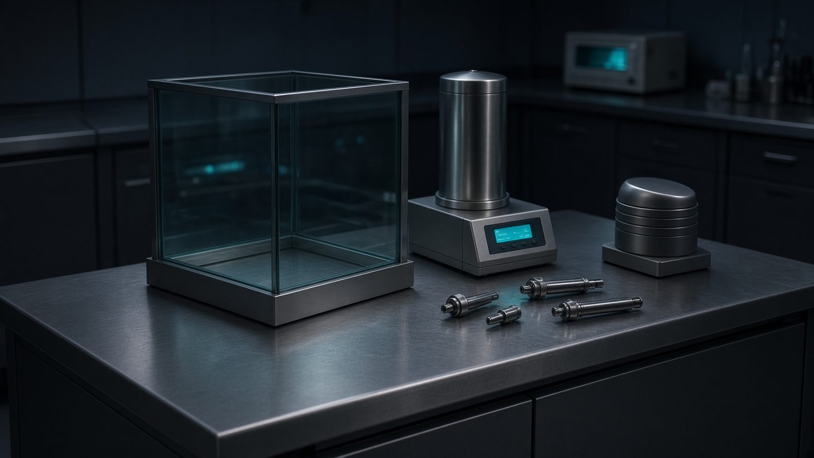 Hot labGenerator, dose calibrator, and L-block — activity, breakthrough, Marinelli.12 eq.
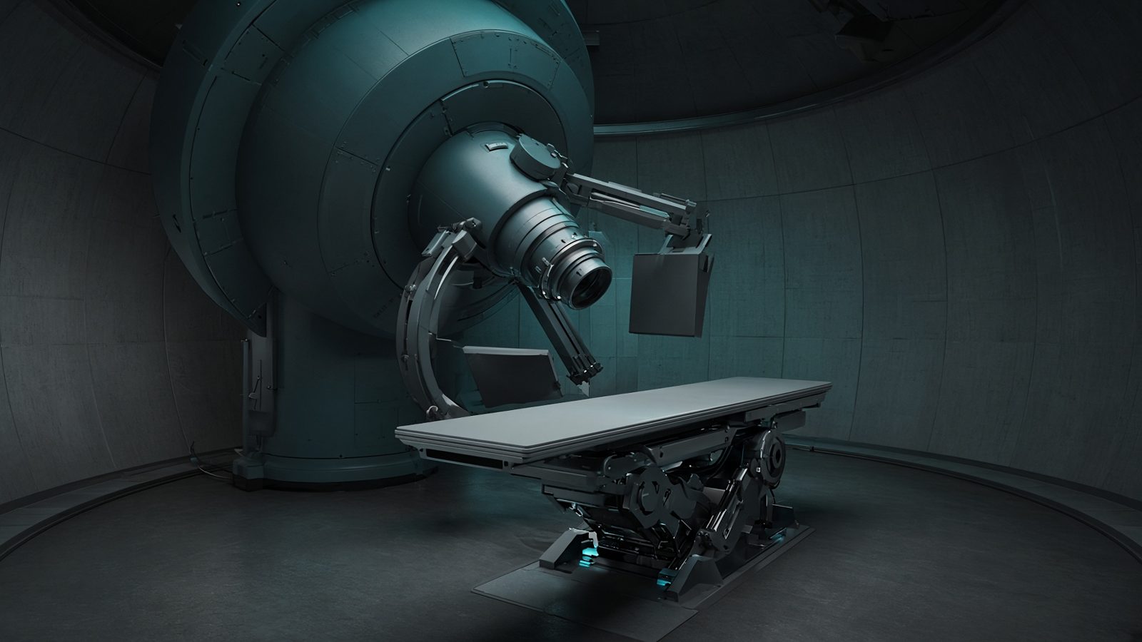 Proton gantryNozzle and snout — residual range, RBE 1.1, and the Bragg peak in the patient.5 eq.