02 Imaging
K-edge energy
K-edge ≈ 1.17 × Moseley Kα. Iodine 33.2 keV, barium 37.4, gadolinium 50.2, tungsten 69.5.
Listen
Listen · English
Simulation
K-edge energy — Change the numbers; the scene follows.
Where it works
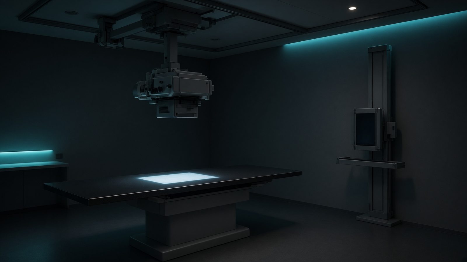
Radiography room
Formula
Typical values
Variables
Results
E_K est.
Estimated K-edge
49.0325keV
E_K
Measured K-edge if known
33.17keV
Explanation
What it means
The K-edge is the binding energy of the K-shell: photoelectric absorption jumps by a factor of 3–5 just above it. Iodine (33.2 keV) and barium (37.4 keV) are matched to the 60–80 kVp spectra of fluoroscopy and GI work. Gadolinium K-edge (50.2 keV) is why Gd can be a CT contrast when iodine is contraindicated. Tungsten K-edge (69.5 keV) is why a 70 kVp beam barely makes W characteristic x-rays. This is a working relation in Diagnostic imaging.
Where it is used
Clinically it sits on the Radiography room — X-ray tube. In the beam filtration and contrast agent — K-edge energy versus kVp. Diagnostic equations live on the tube, the detector, and the patient: magnification, air kerma, CTDI, and why bone lights up at 70 kV. They turn a technique chart into physics you can defend.
Radiography room · Open this machineHow to use it
Enter Z. The calculator uses a hydrogen-like estimate and also lists the textbook measured K-edge when Z matches a common radiology element. Change one input and watch the curve and the simulation follow.
Symbols
- ZAtomic number53
Worked example
A typical case from the default values: Z = 53 (Atomic number). Substituting into the relation gives E_K est. = 49.0325 keV; E_K = 33.17 keV. These are teaching numbers — align them with your machine.
Typical values give
- E_K est. = 49.0325keV
- E_K = 33.17keV
Where it comes from
The displayed formula is the working relation. K-edge ≈ 1.17 × Moseley Kα. Iodine 33.2 keV, barium 37.4, gadolinium 50.2, tungsten 69.5. Usual reference: Bushberg / NIST. Derive it in the specialty lesson, then return here to pin the numbers.
Reference: Bushberg / NIST
Assumptions & limits
Moseley-based estimate, ~5–10% low for high Z. Use NIST XCOM for the real edge. L-edges matter in mammography (Mo L, Rh L) and are not computed here.
Pitfalls
kVp is not the same as effective energy. CTDI is not patient dose — SSDE and organ dose come after. Do not quote DLP as if it were effective dose without a k-factor. Moseley-based estimate, ~5–10% low for high Z. Use NIST XCOM for the real edge. L-edges matter in mammography (Mo L, Rh L) and are not computed here.
Keep this
Technique is physics: kV sets contrast, mAs sets noise, filtration sets the spectrum. Moseley-based estimate, ~5–10% low for high Z. Use NIST XCOM for the real edge. L-edges matter in mammography (Mo L, Rh L) and are not computed here.
In this specialty