02 Imaging
Subject contrast
Fractional difference in transmitted intensity between two regions.
Listen
Listen · English
Simulation
Subject contrast — Change the numbers; the scene follows.
Where it works
Radiography room
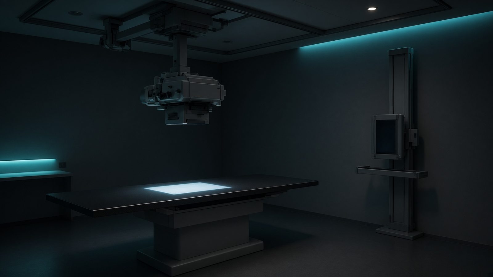
Patient
At the patient entrance — skin dose, subject contrast, photoelectric absorption in tissue.
Open this machineFormula
Variables
Results
C
Contrast
-0.15
C
Contrast
-15%
C_mod
Michelson contrast
-0.1622
Explanation
What it means
Subject contrast is the relative difference in transmitted fluence between two regions, before the detector. It is driven by photoelectric Δμ at low kVp and by density differences at high kVp. This is a working relation in Diagnostic imaging.
Where it is used
Clinically it sits on the Radiography room — Patient. At the patient entrance — skin dose, subject contrast, photoelectric absorption in tissue. Diagnostic equations live on the tube, the detector, and the patient: magnification, air kerma, CTDI, and why bone lights up at 70 kV. They turn a technique chart into physics you can defend.
Radiography room · Open this machineHow to use it
Enter two intensities (or compute them from μ and thickness). Michelson contrast (I2−I1)/((I1+I2)/2) is bounded ±1 and is common in imaging science. Change one input and watch the curve and the simulation follow.
Symbols
- I₁Intensity region 1100 a.u.
- I₂Intensity region 285 a.u.
Worked example
A typical case from the default values: I₁ = 100 a.u. (Intensity region 1); I₂ = 85 a.u. (Intensity region 2). Substituting into the relation gives C = -0.15; C = -15 %; C_mod = -0.1622. These are teaching numbers — align them with your machine.
Typical values give
- C = -0.15
- C = -15%
- C_mod = -0.1622
Where it comes from
The displayed formula is the working relation. Fractional difference in transmitted intensity between two regions. Usual reference: Bushberg. Derive it in the specialty lesson, then return here to pin the numbers.
Reference: Bushberg
Assumptions & limits
This is subject contrast, not displayed (window/level) contrast or detector contrast. Scatter reaching the detector reduces it by 1/(1+SPR).
Pitfalls
kVp is not the same as effective energy. CTDI is not patient dose — SSDE and organ dose come after. Do not quote DLP as if it were effective dose without a k-factor. This is subject contrast, not displayed (window/level) contrast or detector contrast. Scatter reaching the detector reduces it by 1/(1+SPR).
Keep this
Technique is physics: kV sets contrast, mAs sets noise, filtration sets the spectrum. This is subject contrast, not displayed (window/level) contrast or detector contrast. Scatter reaching the detector reduces it by 1/(1+SPR).
In this specialty