02 Imaging
Weighted and volumetric CTDI
CTDI_w and CTDI_vol from center, periphery, and pitch.
Listen
Listen · English
Simulation
Weighted and volumetric CTDI — Change the numbers; the scene follows.
Where it works
CT scanner
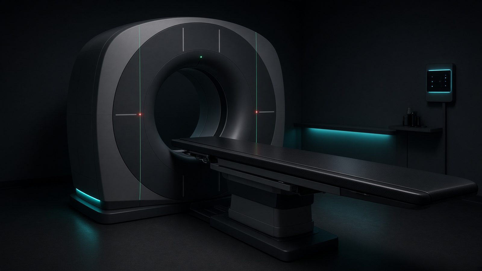
Gantry
Inside the rotating gantry: tube, bowtie, and detectors that define CTDI and HU.
Open this machineFormula
Variables
Results
CTDI_w
Weighted CTDI
10.6667mGy
CTDI_vol
Volumetric CTDI
10.6667mGy
Explanation
What it means
CTDI_w weights the centre and periphery of a CTDI phantom (1/3 centre + 2/3 periphery) to represent average dose in the slice. Dividing by pitch gives CTDI_vol, the standard dose index displayed on the console. This is a working relation in Diagnostic imaging.
Where it is used
Clinically it sits on the CT scanner — Gantry. Inside the rotating gantry: tube, bowtie, and detectors that define CTDI and HU. Diagnostic equations live on the tube, the detector, and the patient: magnification, air kerma, CTDI, and why bone lights up at 70 kV. They turn a technique chart into physics you can defend.
CT scanner · Open this machineHow to use it
Use 16 cm phantom for head, 32 cm for body. Pitch > 1 lowers CTDI_vol; pitch < 1 (overlapping) raises it. CTDI_vol × scan length = DLP. Change one input and watch the curve and the simulation follow.
Symbols
- CTDI_cCenter CTDI8 mGy
- CTDI_pPeriphery CTDI12 mGy
- pitchPitch1
Worked example
A typical case from the default values: CTDI_c = 8 mGy (Center CTDI); CTDI_p = 12 mGy (Periphery CTDI); pitch = 1 (Pitch). Substituting into the relation gives CTDI_w = 10.6667 mGy; CTDI_vol = 10.6667 mGy. These are teaching numbers — align them with your machine.
Typical values give
- CTDI_w = 10.6667mGy
- CTDI_vol = 10.6667mGy
Where it comes from
The displayed formula is the working relation. CTDI_w and CTDI_vol from center, periphery, and pitch. Usual reference: AAPM / IEC CT dosimetry. Derive it in the specialty lesson, then return here to pin the numbers.
Reference: AAPM / IEC CT dosimetry
Assumptions & limits
Phantom indices, not patient dose. Size-specific dose estimate (SSDE) applies a conversion from effective diameter. Direct-beam only — overscan is extra.
Pitfalls
kVp is not the same as effective energy. CTDI is not patient dose — SSDE and organ dose come after. Do not quote DLP as if it were effective dose without a k-factor. Phantom indices, not patient dose. Size-specific dose estimate (SSDE) applies a conversion from effective diameter. Direct-beam only — overscan is extra.
Keep this
Technique is physics: kV sets contrast, mAs sets noise, filtration sets the spectrum. Phantom indices, not patient dose. Size-specific dose estimate (SSDE) applies a conversion from effective diameter. Direct-beam only — overscan is extra.
In this specialty