02 Imaging
Mean glandular dose (Dance)
AGD = K g c s. Incident air kerma × conversion factors for thickness, composition, spectrum.
Listen
Listen · English
Simulation
Mean glandular dose (Dance) — Change the numbers; the scene follows.
Where it works
Mammography unit
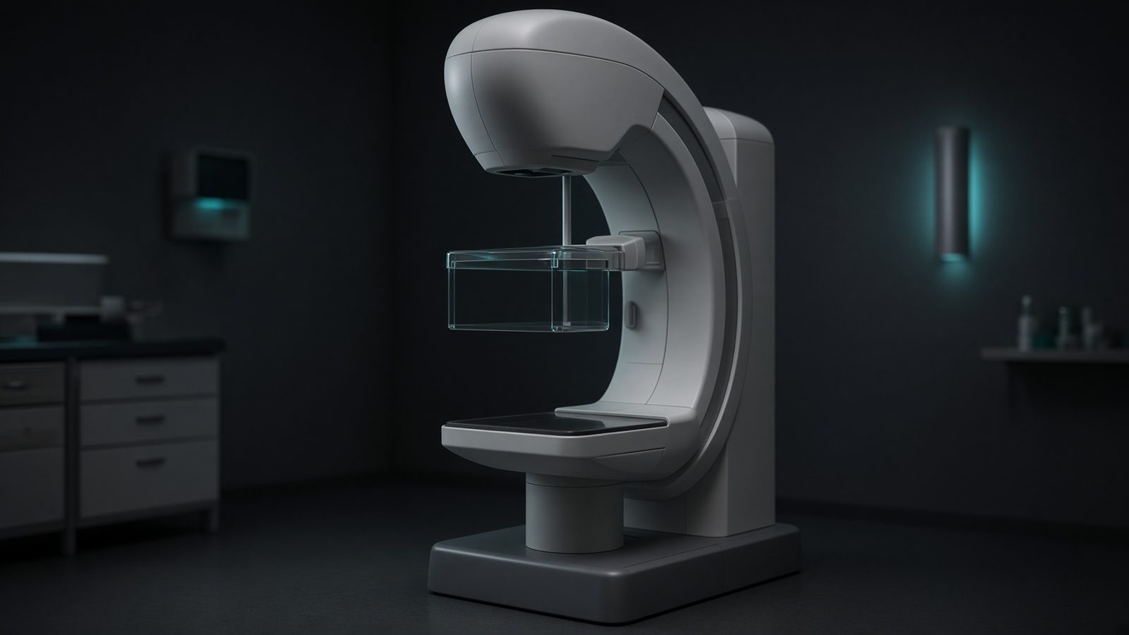
Breast / detector
In the compressed breast, just above the detector — average glandular dose.
Open this machineFormula
Typical values
Variables
Results
AGD
Mean glandular dose
1.464mGy
Explanation
What it means
European and UK mammography dose is mean glandular dose, not entrance air kerma. Dance factored the conversion into g (glandularity 50%, HVL and thickness), c (composition other than 50%) and s (spectrum: Mo/Mo = 1, Mo/Rh, W/Rh, W/Ag, W/Al). Typical screening AGD is 1–2 mGy per view; EUREF achievable is < 2.0 mGy at 4.5 cm 50% glandularity. This is a working relation in Diagnostic imaging.
Where it is used
Clinically it sits on the Mammography unit — Breast / detector. In the compressed breast, just above the detector — average glandular dose. Diagnostic equations live on the tube, the detector, and the patient: magnification, air kerma, CTDI, and why bone lights up at 70 kV. They turn a technique chart into physics you can defend.
Mammography unit · Open this machineHow to use it
Enter incident air kerma K (mGy) at the upper surface without backscatter, then g, c, s from Dance tables for your HVL, thickness and anode/filter. Default g=0.183 (5 cm, HVL 0.35 mm Al), c=1, s=1 (Mo/Mo). Change one input and watch the curve and the simulation follow.
Symbols
- KIncident air kerma8 mGy
- gDance g0.183
- cComposition c1
- sSpectrum s1
Worked example
A typical case from the default values: K = 8 mGy (Incident air kerma); g = 0.183 (Dance g); c = 1 (Composition c); s = 1 (Spectrum s). Substituting into the relation gives AGD = 1.464 mGy. These are teaching numbers — align them with your machine.
Typical values give
- AGD = 1.464mGy
Where it comes from
The displayed formula is the working relation. AGD = K g c s. Incident air kerma × conversion factors for thickness, composition, spectrum. Usual reference: Dance 1990 / 2000 / EUREF. Derive it in the specialty lesson, then return here to pin the numbers.
Reference: Dance 1990 / 2000 / EUREF
Assumptions & limits
Does not look up Dance tables — you must supply g, c, s. Wu / Boone American methods use a different factorisation. K must be incident air kerma, not including backscatter (ESAKs that include BSF need dividing by BSF ≈ 1.1).
Pitfalls
kVp is not the same as effective energy. CTDI is not patient dose — SSDE and organ dose come after. Do not quote DLP as if it were effective dose without a k-factor. Does not look up Dance tables — you must supply g, c, s. Wu / Boone American methods use a different factorisation. K must be incident air kerma, not including backscatter (ESAKs that include BSF need dividing by BSF ≈ 1.1).
Keep this
Technique is physics: kV sets contrast, mAs sets noise, filtration sets the spectrum. Does not look up Dance tables — you must supply g, c, s. Wu / Boone American methods use a different factorisation. K must be incident air kerma, not including backscatter (ESAKs that include BSF need dividing by BSF ≈ 1.1).
In this specialty