03 Nuclear
Attenuation correction factor
PET: ACF = exp(∫ μ ds) along the LOR. SPECT: exp(μ d) for a broad-beam μ.
Listen
Listen · English
Simulation
Attenuation correction factor — Change the numbers; the scene follows.
Where it works
PET/CT
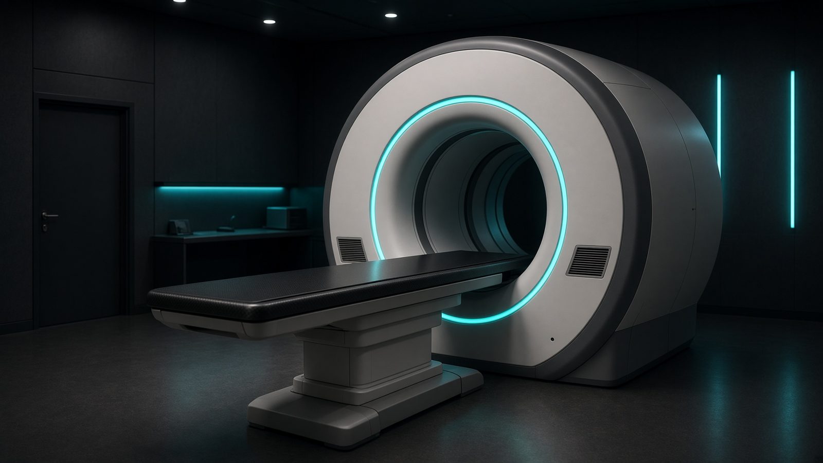
CT component
On the CT component of the hybrid gantry — µ-map for attenuation correction.
Open this machineFormula
Typical values
Variables
Results
ACF
Attenuation correction factor
17.8143
e^{−μd}
Transmission
0.0561
Curve
Explanation
What it means
A 30-cm abdomen at 511 keV (μ ≈ 0.096 cm⁻¹) attenuates a coincidence by e^{μ D} ≈ 18 — that is why uncorrected PET looks skin-bright and liver-dark. CT-based AC measures μ at ~60–80 keV and bilinearly scales it to 511 keV (or to the SPECT window). A stale μ-map (misregistration, truncation, metal) is the most common AC artefact. This is a working relation in Nuclear medicine.
Where it is used
Clinically it sits on the PET/CT — CT component. On the CT component of the hybrid gantry — µ-map for attenuation correction. Nuclear-medicine relations sit in the hot lab, on the camera, and in the voxel: decay, SUV, TOF, and counting statistics. They decide whether an uptake is real or a clock error.
PET/CT · Open this machineHow to use it
Enter μ (cm⁻¹) and path length d (cm). For PET coincidences d is the full LOR through the patient; for SPECT d is source-to-skin along the projection. Default μ = 0.096 (511 keV water) or 0.15 (140 keV water). Change one input and watch the curve and the simulation follow.
Symbols
- μLinear attenuation0.096 cm⁻¹
- dPath length30 cm
Worked example
A typical case from the default values: μ = 0.096 cm⁻¹ (Linear attenuation); d = 30 cm (Path length). Substituting into the relation gives ACF = 17.8143; e^{−μd} = 0.0561. These are teaching numbers — align them with your machine.
Typical values give
- ACF = 17.8143
- e^{−μd} = 0.0561
Where it comes from
The displayed formula is the working relation. PET: ACF = exp(∫ μ ds) along the LOR. SPECT: exp(μ d) for a broad-beam μ. Usual reference: Cherry / Bailey. Derive it in the specialty lesson, then return here to pin the numbers.
Reference: Cherry / Bailey
Assumptions & limits
Uniform μ, no scatter, no bed, no truncation. Broad-beam SPECT μ is smaller than narrow-beam. Does not replace a CT μ-map.
Pitfalls
Activity is not counts. SUV needs the true injected activity, the residual, and the correct decay time — a clock off by 10 min on ¹⁸F is a several-percent error. Do not compare SUVs across reconstructions. Uniform μ, no scatter, no bed, no truncation. Broad-beam SPECT μ is smaller than narrow-beam. Does not replace a CT μ-map.
Keep this
Write the assay time next to every activity. Decay does the rest. Uniform μ, no scatter, no bed, no truncation. Broad-beam SPECT μ is smaller than narrow-beam. Does not replace a CT μ-map.
In this specialty