03 Nuclear
PET time-of-flight localisation
Δx = c Δt / 2. A 400 ps coincidence window localises the annihilation to ~6 cm FWHM.
Listen
Listen · English
Simulation
PET time-of-flight localisation — Change the numbers; the scene follows.
Where it works
PET/CT
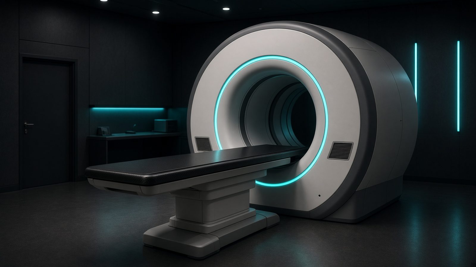
PET detectors
In the PET detector ring — coincidence timing, noise-equivalent counts, randoms.
Open this machineFormula
Variables
Results
Δx
TOF localisation
5.9958cm
Δx
TOF localisation
59.9585mm
Curve
Explanation
What it means
In PET the two 511 keV photons are born together. The time difference Δt between their arrivals encodes the position along the line of response: the source is closer to the earlier crystal by Δx = c Δt / 2. Conventional PET (Δt ~ 2–4 ns) only tells you the LOR; TOF PET (200–400 ps) shrinks the uncertainty to a few centimetres and gains SNR roughly as √(D / Δx) where D is the patient diameter. This is a working relation in Nuclear medicine.
Where it is used
Clinically it sits on the PET/CT — PET detectors. In the PET detector ring — coincidence timing, noise-equivalent counts, randoms. Nuclear-medicine relations sit in the hot lab, on the camera, and in the voxel: decay, SUV, TOF, and counting statistics. They decide whether an uptake is real or a clock error.
PET/CT · Open this machineHow to use it
Enter timing resolution FWHM in picoseconds. 500 ps → 7.5 cm; 200 ps → 3.0 cm; 60 ps (research LaBr / Cherenkov) → 9 mm. Pair it with the SNR-gain calculator mentally: gain ≈ √(30 cm / Δx). Change one input and watch the curve and the simulation follow.
Symbols
- ΔtTiming FWHM400 ps
Worked example
A typical case from the default values: Δt = 400 ps (Timing FWHM). Substituting into the relation gives Δx = 5.9958 cm; Δx = 59.9585 mm. These are teaching numbers — align them with your machine.
Typical values give
- Δx = 5.9958cm
- Δx = 59.9585mm
Where it comes from
Photon 1 travels x, photon 2 travels L−x. Δt = (x − (L−x))/c = (2x − L)/c, so x − L/2 = c Δt / 2.
Reference: Cherry, Sorenson & Phelps / Surti
Assumptions & limits
Vacuum speed of light; refractive index in crystal is already folded into the published Δt of the scanner. Does not include DOI parallax or the reconstruction kernel, which further blur Δx.
Pitfalls
Activity is not counts. SUV needs the true injected activity, the residual, and the correct decay time — a clock off by 10 min on ¹⁸F is a several-percent error. Do not compare SUVs across reconstructions. Vacuum speed of light; refractive index in crystal is already folded into the published Δt of the scanner. Does not include DOI parallax or the reconstruction kernel, which further blur Δx.
Keep this
Write the assay time next to every activity. Decay does the rest. Vacuum speed of light; refractive index in crystal is already folded into the published Δt of the scanner. Does not include DOI parallax or the reconstruction kernel, which further blur Δx.
In this specialty