01 Therapy
van Herk PTV margin
M = 2.5 Σ + 0.7 σ so that 90% of patients get ≥95% dose to the CTV.
Listen
Listen · English
Simulation
van Herk PTV margin — Change the numbers; the scene follows.
Where it works
Linear accelerator
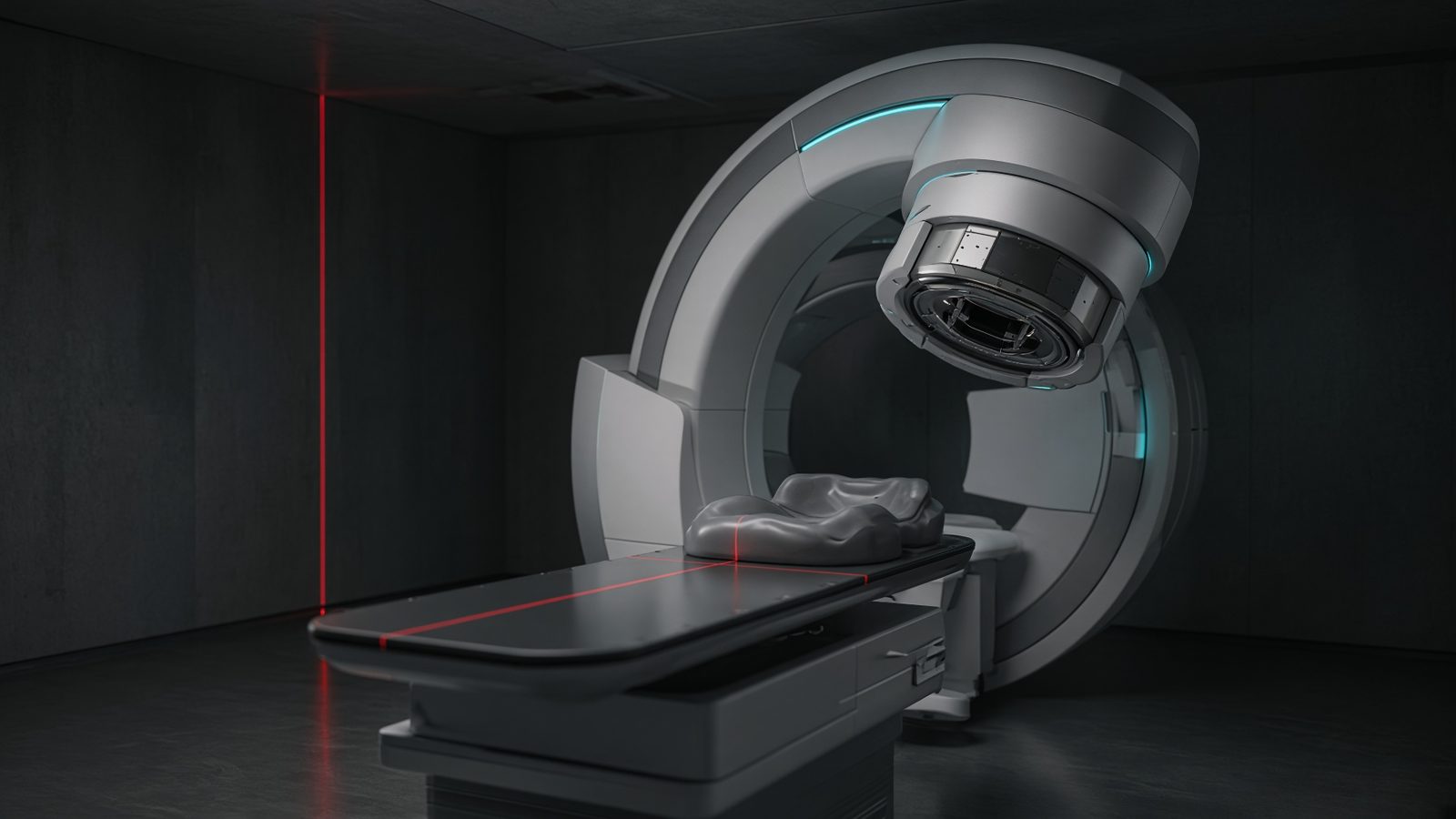
Isocenter
At isocenter, on the central axis through the patient (or a phantom in the same place).
Open this machineFormula
Variables
Results
M
PTV margin
7.1mm
2.5 Σ
Systematic term
5mm
0.7 σ
Random term
2.1mm
Curve
Explanation
What it means
Systematic error Σ (preparation: delineation, setup bias, image registration) shifts the whole treatment and must be covered generously (2.5 Σ ≈ 90% of the population). Random error σ (daily setup, organ motion) blurs the dose and is covered by 0.7 σ, which restores the 95% isodose to the CTV. Add a small penumbra term 1.64 σ_p if you start from a 50% instead of 95% edge — omitted here. This is a working relation in Radiotherapy.
Where it is used
Clinically it sits on the Linear accelerator — Isocenter. At isocenter, on the central axis through the patient (or a phantom in the same place). Radiotherapy equations sit at the console and in the bunker: output, depth dose, equivalent square, and the monitor units that treat the patient. Hand-calc them beside the TPS, never instead of a commissioned plan.
Linear accelerator · Open this machineHow to use it
Enter RMS systematic Σ and RMS random σ in mm (combine sources in quadrature beforehand: Σ = √(Σ_setup²+Σ_delin²+…). A typical image-guided prostate: Σ 2 mm, σ 3 mm → M ≈ 7.1 mm. Change one input and watch the curve and the simulation follow.
Symbols
- ΣSystematic RMS2 mm
- σRandom RMS3 mm
Worked example
A typical case from the default values: Σ = 2 mm (Systematic RMS); σ = 3 mm (Random RMS). Substituting into the relation gives M = 7.1 mm; 2.5 Σ = 5 mm; 0.7 σ = 2.1 mm. These are teaching numbers — align them with your machine.
Typical values give
- M = 7.1mm
- 2.5 Σ = 5mm
- 0.7 σ = 2.1mm
Where it comes from
Population coverage: the systematic shift is Gaussian, and 2.5 Σ is the 90% quantile of the 3-D vector (actually 2.5 is for 90% of patients in all directions combined). The 0.7 σ term is the extra margin that moves the 95% isodose back onto the CTV after Gaussian blurring of a typical penumbra.
Reference: van Herk et al., IJROBP 2000
Assumptions & limits
Linear approximation for a spherical CTV, perfect 95% conformal dose, no rotation or deformation. SBRT with a steep gradient and hypofractionation needs a recipe that also looks at number of fractions. Delineation uncertainty is often the hidden dominant Σ.
Pitfalls
Never mix PDD from one SSD with TMR from another without converting. Field size at the surface is not the size at isocentre. A hand MU is a check, not a treatment. Linear approximation for a spherical CTV, perfect 95% conformal dose, no rotation or deformation. SBRT with a steep gradient and hypofractionation needs a recipe that also looks at number of fractions. Delineation uncertainty is often the hidden dominant Σ.
Keep this
Name the SSD, energy, and field size with every PDD or TMR you quote. Linear approximation for a spherical CTV, perfect 95% conformal dose, no rotation or deformation. SBRT with a steep gradient and hypofractionation needs a recipe that also looks at number of fractions. Delineation uncertainty is often the hidden dominant Σ.
In this specialty