08 Ultrasound
Thermal index
TI = W₀ / W_deg, the output power divided by the power that raises tissue by 1 °C.
Listen
Listen · English
Simulation
Thermal index — Change the numbers; the scene follows.
Where it works
Ultrasound
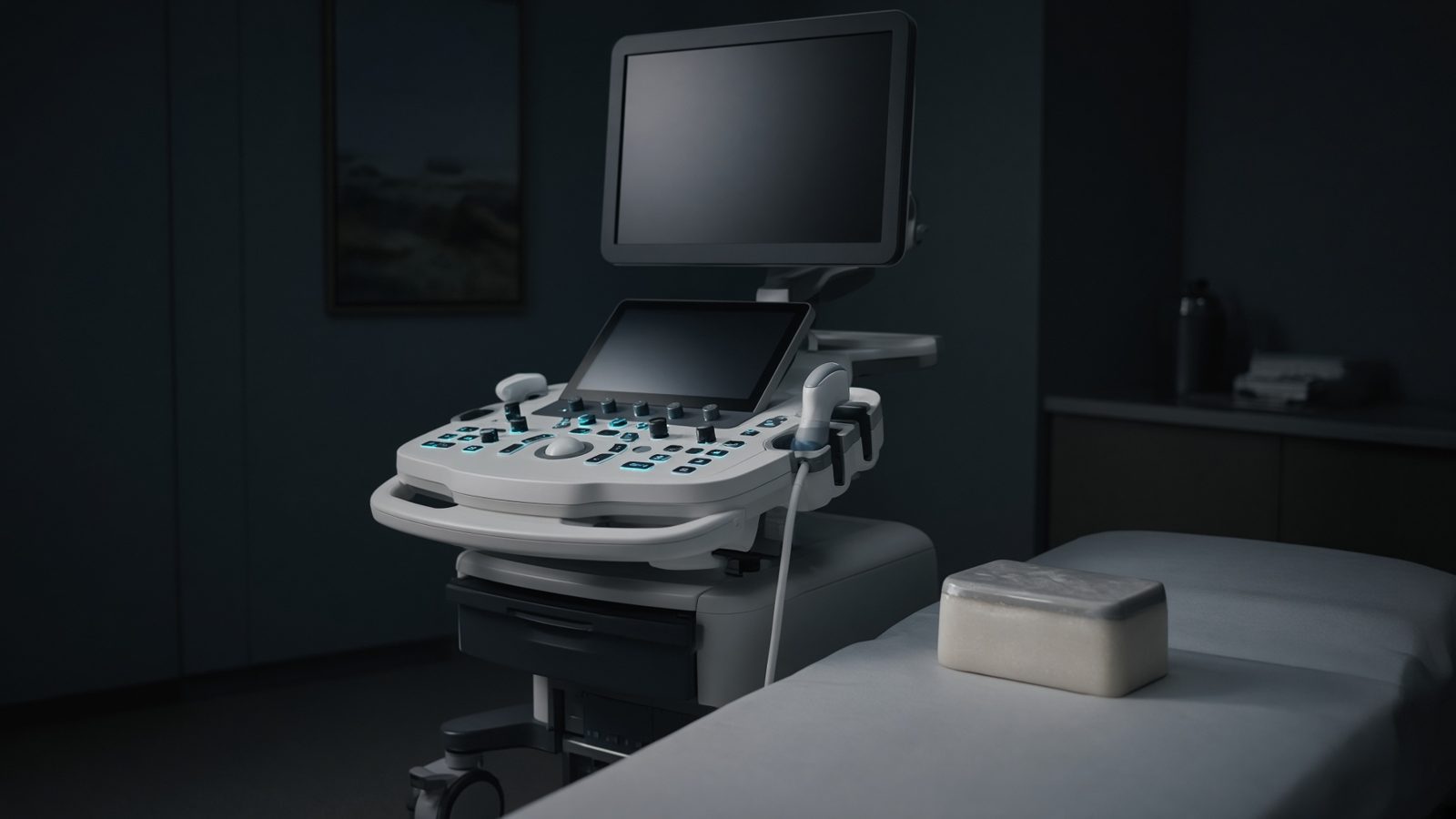
Tissue path
Along the beam in tissue — impedance mismatch, Doppler shift, MI/TI at the focus.
Open this machineFormula
Variables
Results
TI
Thermal index
0.8
Explanation
What it means
Thermal index is an on-screen ‘how many degrees might this heat the tissue’ under a standardised model: TIS (soft tissue), TIB (bone at focus), TIC (cranial bone at surface). TI = 1 means the model predicts ~1 °C at the worst-case point. Obstetric practice keeps TI ≤ 0.7 for long surveys and uses TIB once bone is ossified. It is not a thermometer. This is a working relation in Ultrasound.
Where it is used
Clinically it sits on the Ultrasound — Tissue path. Along the beam in tissue — impedance mismatch, Doppler shift, MI/TI at the focus. Ultrasound equations sit on the probe face and along the beam: impedance, Snell, Doppler, MI and TI. They explain why gel matters, why aliasing appears, and why a mechanical index is on the screen.
Ultrasound · Open this machineHow to use it
Enter output power W₀ (mW) and the 1-°C power W_deg (mW) from the system’s ODS model. If you only have a displayed TI, this calculator will not invert it — it is the definition itself. Default 40 mW / 50 mW → TI = 0.8. Change one input and watch the curve and the simulation follow.
Symbols
- W_0Output power40 mW
- W_degPower for +1 °C50 mW
Worked example
A typical case from the default values: W_0 = 40 mW (Output power); W_deg = 50 mW (Power for +1 °C). Substituting into the relation gives TI = 0.8. These are teaching numbers — align them with your machine.
Typical values give
- TI = 0.8
Where it comes from
The displayed formula is the working relation. TI = W₀ / W_deg, the output power divided by the power that raises tissue by 1 °C. Usual reference: AIUM / NEMA ODS / IEC 62359. Derive it in the specialty lesson, then return here to pin the numbers.
Reference: AIUM / NEMA ODS / IEC 62359
Assumptions & limits
The hard part is W_deg, which depends on path, absorption, perfusion and whether bone is in the focus — the scanner estimates it. This is the definition, not a finite-element temperature map. Exposure time still matters (a TI of 1 for 30 s is not a TI of 1 for 30 min).
Pitfalls
Soft-tissue 1540 m/s is an assumption — not a measurement in that patient. Doppler angle 90° gives no shift. MI and TI are on-screen estimates, not absorbed dose. The hard part is W_deg, which depends on path, absorption, perfusion and whether bone is in the focus — the scanner estimates it. This is the definition, not a finite-element temperature map. Exposure time still matters (a TI of 1 for 30 s is not a TI of 1 for 30 min).
Keep this
Gel, angle, and assumed speed of sound — get those three right before you trust a centimetre. The hard part is W_deg, which depends on path, absorption, perfusion and whether bone is in the focus — the scanner estimates it. This is the definition, not a finite-element temperature map. Exposure time still matters (a TI of 1 for 30 s is not a TI of 1 for 30 min).
In this specialty