08 Ultrasound
Simplified Bernoulli pressure drop
ΔP (mmHg) = 4 v² with v in m/s. Peak CW-Doppler velocity across a valve.
Listen
Listen · English
Simulation
Simplified Bernoulli pressure drop — Change the numbers; the scene follows.
Where it works
Ultrasound
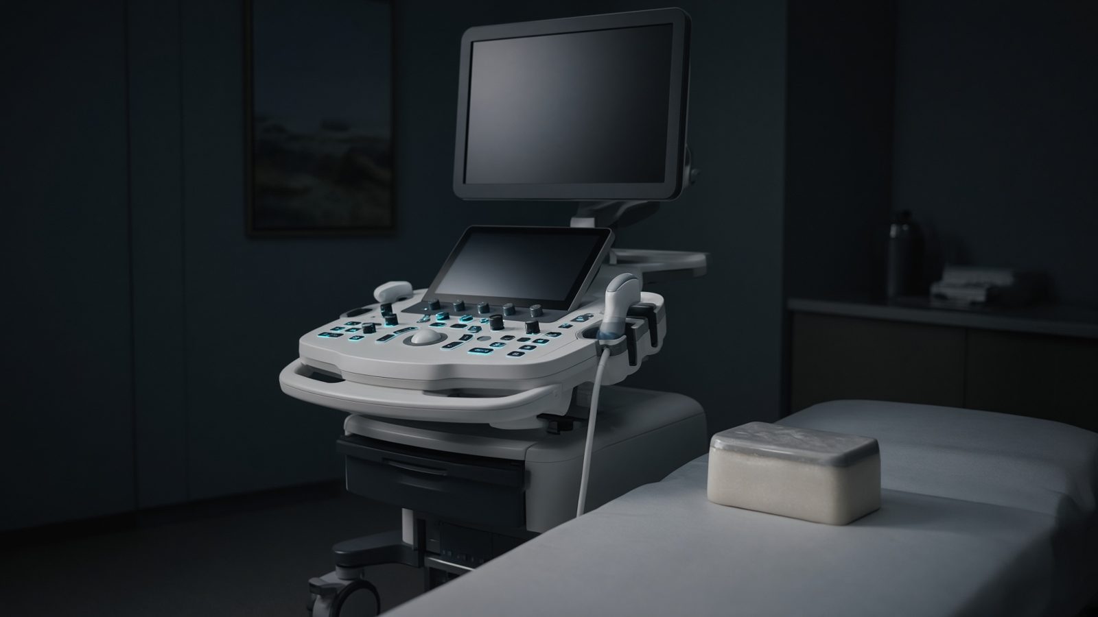
Tissue path
Along the beam in tissue — impedance mismatch, Doppler shift, MI/TI at the focus.
Open this machineFormula
Variables
Results
ΔP_4v²
Simplified
64mmHg
ΔP
With proximal velocity
64mmHg
Curve
Explanation
What it means
Starting from Bernoulli’s ½ ρ (v₂² − v₁²) and ρ_blood ≈ 1060 kg/m³, converting to mmHg and dropping the proximal velocity v₁ (and flow acceleration / resistance), cardiology obtains the mnemonic ΔP = 4 v². A 4 m/s CW jet is a 64 mmHg gradient — severe aortic stenosis if that is the peak transvalvular velocity. Add proximal velocity as 4(v₂² − v₁²) when v₁ is not negligible (LVOT > 1.5 m/s). This is a working relation in Ultrasound.
Where it is used
Clinically it sits on the Ultrasound — Tissue path. Along the beam in tissue — impedance mismatch, Doppler shift, MI/TI at the focus. Ultrasound equations sit on the probe face and along the beam: impedance, Snell, Doppler, MI and TI. They explain why gel matters, why aliasing appears, and why a mechanical index is on the screen.
Ultrasound · Open this machineHow to use it
Enter jet velocity v₂ (m/s) and optionally proximal v₁. 4 m/s / 0.9 m/s → 4(16 − 0.81) = 60.8 mmHg versus 64 mmHg simplified. Change one input and watch the curve and the simulation follow.
Symbols
- v_2Jet velocity4 m/s
- v_1Proximal velocity0 m/s
Worked example
A typical case from the default values: v_2 = 4 m/s (Jet velocity); v_1 = 0 m/s (Proximal velocity). Substituting into the relation gives ΔP_4v² = 64 mmHg; ΔP = 64 mmHg. These are teaching numbers — align them with your machine.
Typical values give
- ΔP_4v² = 64mmHg
- ΔP = 64mmHg
Where it comes from
The displayed formula is the working relation. ΔP (mmHg) = 4 v² with v in m/s. Peak CW-Doppler velocity across a valve. Usual reference: Hatle / Otto (echo) / Bernoulli. Derive it in the specialty lesson, then return here to pin the numbers.
Reference: Hatle / Otto (echo) / Bernoulli
Assumptions & limits
Inviscid, aligned CW beam (angle = 0), peak instantaneous (not mean) gradient. Mean gradient needs tracing the VTI. Pressure recovery distal to a stenosis can make the Doppler gradient overestimate the catheter gradient.
Pitfalls
Soft-tissue 1540 m/s is an assumption — not a measurement in that patient. Doppler angle 90° gives no shift. MI and TI are on-screen estimates, not absorbed dose. Inviscid, aligned CW beam (angle = 0), peak instantaneous (not mean) gradient. Mean gradient needs tracing the VTI. Pressure recovery distal to a stenosis can make the Doppler gradient overestimate the catheter gradient.
Keep this
Gel, angle, and assumed speed of sound — get those three right before you trust a centimetre. Inviscid, aligned CW beam (angle = 0), peak instantaneous (not mean) gradient. Mean gradient needs tracing the VTI. Pressure recovery distal to a stenosis can make the Doppler gradient overestimate the catheter gradient.
In this specialty