08 Ultrasound
Doppler shift
Frequency shift from flow speed and probe angle.
Listen
Listen · English
Simulation
Doppler shift — Change the numbers; the scene follows.
Where it works
Ultrasound
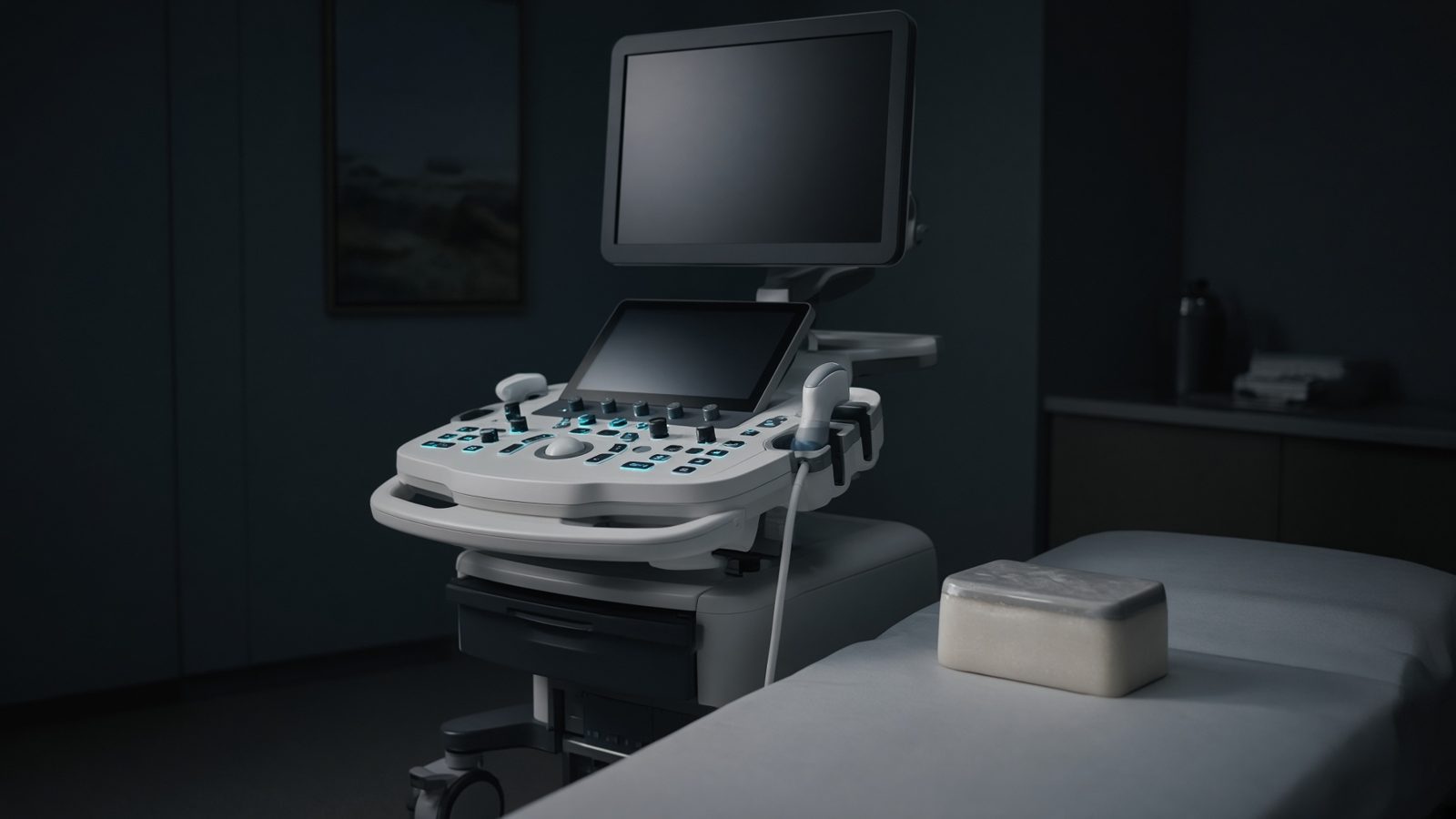
Tissue path
Along the beam in tissue — impedance mismatch, Doppler shift, MI/TI at the focus.
Open this machineFormula
Variables
Results
Δf
Doppler shift
1,623.4Hz
Δf
Doppler shift
1.6234kHz
Curve
Explanation
What it means
The Doppler shift from flowing blood is Δf = 2 v f₀ cosθ / c. The 2 is transmit plus receive. At 90° the shift is zero — angle correct or don’t quote velocity. Alias when |Δf| > PRF/2. This is a working relation in Ultrasound.
Where it is used
Clinically it sits on the Ultrasound — Tissue path. Along the beam in tissue — impedance mismatch, Doppler shift, MI/TI at the focus. Ultrasound equations sit on the probe face and along the beam: impedance, Snell, Doppler, MI and TI. They explain why gel matters, why aliasing appears, and why a mechanical index is on the screen.
Ultrasound · Open this machineHow to use it
v in cm/s, f₀ in MHz, θ between beam and flow. The curve vs angle shows why 60° is a common compromise (error grows fast beyond that). Change one input and watch the curve and the simulation follow.
Symbols
- vFlow speed50 cm/s
- f₀Probe frequency5 MHz
- θAngle60 °
- cSpeed of sound1,540 m/s
Worked example
A typical case from the default values: v = 50 cm/s (Flow speed); f₀ = 5 MHz (Probe frequency); θ = 60 ° (Angle); c = 1,540 m/s (Speed of sound). Substituting into the relation gives Δf = 1,623.4 Hz; Δf = 1.6234 kHz. These are teaching numbers — align them with your machine.
Typical values give
- Δf = 1,623.4Hz
- Δf = 1.6234kHz
Where it comes from
The displayed formula is the working relation. Frequency shift from flow speed and probe angle. Usual reference: Bushberg / Edelman. Derive it in the specialty lesson, then return here to pin the numbers.
Reference: Bushberg / Edelman
Assumptions & limits
Single scatterer, constant v, no intrinsic spectral broadening. Stenotic jets are not a single v. High-PRF / CW modes relax the aliasing limit differently.
Pitfalls
Soft-tissue 1540 m/s is an assumption — not a measurement in that patient. Doppler angle 90° gives no shift. MI and TI are on-screen estimates, not absorbed dose. Single scatterer, constant v, no intrinsic spectral broadening. Stenotic jets are not a single v. High-PRF / CW modes relax the aliasing limit differently.
Keep this
Gel, angle, and assumed speed of sound — get those three right before you trust a centimetre. Single scatterer, constant v, no intrinsic spectral broadening. Stenotic jets are not a single v. High-PRF / CW modes relax the aliasing limit differently.
In this specialty