08 Ultrasound
Lateral resolution
Beam width ≈ 1.2 λ z / D at the focus (Airy / diffraction).
Listen
Listen · English
Simulation
Lateral resolution — Change the numbers; the scene follows.
Where it works
Ultrasound
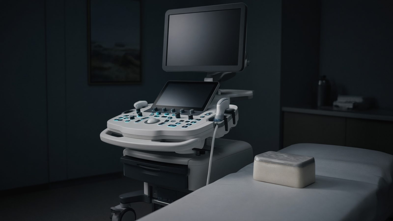
QA phantom
On the tissue-mimicking phantom — axial and lateral resolution are checked here.
Open this machineFormula
Variables
Results
λ
Wavelength
0.308mm
w
Lateral resolution
1.1088mm
Explanation
What it means
Lateral resolution is the beam width, roughly 1.2 λ z / D at a focus (diffraction). Deeper, or a smaller probe footprint, widens the beam. Multiple transmit foci and dynamic receive focusing try to keep w small over a range of depths. This is a working relation in Ultrasound.
Where it is used
Clinically it sits on the Ultrasound — QA phantom. On the tissue-mimicking phantom — axial and lateral resolution are checked here. Ultrasound equations sit on the probe face and along the beam: impedance, Snell, Doppler, MI and TI. They explain why gel matters, why aliasing appears, and why a mechanical index is on the screen.
Ultrasound · Open this machineHow to use it
5 MHz, 6 cm, 20 mm aperture → ~0.6 mm. Elevation (slice) thickness is a separate, usually worse, number set by the lens in 1-D arrays. Change one input and watch the curve and the simulation follow.
Symbols
- fFrequency5 MHz
- zDepth / focus6 cm
- DAperture width20 mm
- cSpeed of sound1,540 m/s
Worked example
A typical case from the default values: f = 5 MHz (Frequency); z = 6 cm (Depth / focus); D = 20 mm (Aperture width); c = 1,540 m/s (Speed of sound). Substituting into the relation gives λ = 0.308 mm; w = 1.1088 mm. These are teaching numbers — align them with your machine.
Typical values give
- λ = 0.308mm
- w = 1.1088mm
Where it comes from
The displayed formula is the working relation. Beam width ≈ 1.2 λ z / D at the focus (Airy / diffraction). Usual reference: Bushberg. Derive it in the specialty lesson, then return here to pin the numbers.
Reference: Bushberg
Assumptions & limits
Continuous-wave circular aperture idealisation. Apodisation, grating lobes and the actual f-number of the array change the factor 1.2.
Pitfalls
Soft-tissue 1540 m/s is an assumption — not a measurement in that patient. Doppler angle 90° gives no shift. MI and TI are on-screen estimates, not absorbed dose. Continuous-wave circular aperture idealisation. Apodisation, grating lobes and the actual f-number of the array change the factor 1.2.
Keep this
Gel, angle, and assumed speed of sound — get those three right before you trust a centimetre. Continuous-wave circular aperture idealisation. Apodisation, grating lobes and the actual f-number of the array change the factor 1.2.
In this specialty