06 Dose
MIRD absorbed dose
Target-organ dose from cumulated activity and the S-value.
Listen
Listen · English
Simulation
MIRD absorbed dose — Change the numbers; the scene follows.
Where it works
PET/CT
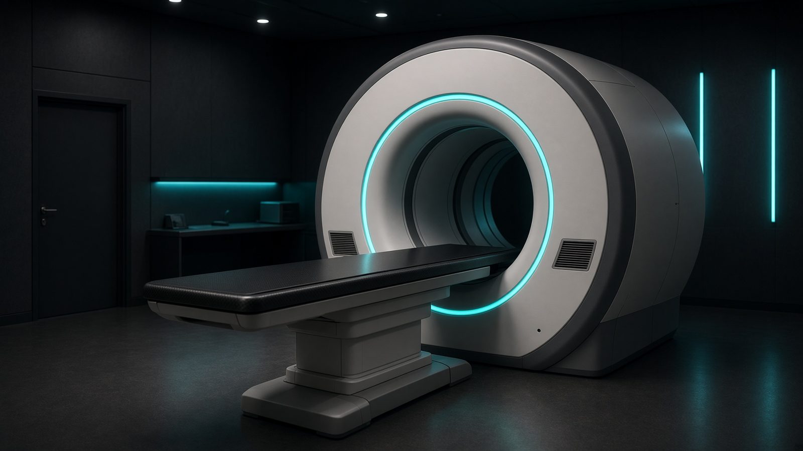
Patient / uptake
In the patient on the PET couch — activity concentration, SUV, and internal dose.
Open this machineFormula
Variables
Results
D
Absorbed dose
1,200mGy
D
Absorbed dose
1.2Gy
Explanation
What it means
MIRD absorbed dose in a target organ is cumulated activity in the source times the S-value (mean dose per nuclear transformation, including self-dose and cross-dose). D = Ã S, with consistent units. This is a working relation in Dosimetry.
Where it is used
Clinically it sits on the PET/CT — Patient / uptake. In the patient on the PET couch — activity concentration, SUV, and internal dose. Dosimetry is the chamber in water under the linac, or the well counter in the hot lab: TG-51, TRS-398, kerma, and recombination. These numbers are the calibration the rest of the department borrows.
PET/CT · Open this machineHow to use it
à from the residence-time / cumulated-activity calculators. S from OLINDA/MIRD pamphlets (mGy/MBq·s or mGy/MBq·h). Sum over all source organs including remainder. Change one input and watch the curve and the simulation follow.
Symbols
- ÃCumulated activity1.0000e+8 MBq·s
- SS-value1.2000e-5 mGy/(MBq·s)
Worked example
A typical case from the default values: Ã = 1.0000e+8 MBq·s (Cumulated activity); S = 1.2000e-5 mGy/(MBq·s) (S-value). Substituting into the relation gives D = 1,200 mGy; D = 1.2 Gy. These are teaching numbers — align them with your machine.
Typical values give
- D = 1,200mGy
- D = 1.2Gy
Where it comes from
The displayed formula is the working relation. Target-organ dose from cumulated activity and the S-value. Usual reference: MIRD primer. Derive it in the specialty lesson, then return here to pin the numbers.
Reference: MIRD primer
Assumptions & limits
Standard phantom S-values, not the patient’s anatomy. Therapy (I-131, Lu-177) should move toward voxel S-values / Monte Carlo when activity is high.
Pitfalls
kQ is for that chamber and that beam quality — not a neighbour's value. Polarity and recombination are measured, not copied. A ⁶⁰Co N_D,w is not an MV calibration until kQ is applied. Standard phantom S-values, not the patient’s anatomy. Therapy (I-131, Lu-177) should move toward voxel S-values / Monte Carlo when activity is high.
Keep this
à in MBq·s and S in mGy/(MBq·s), or keep units consistent.
In this specialty